TOP

HOME


Show of the Month May 2015

Show of the Month May 2 2015

Show of the Month May 9 2015

Show of the Month May 16 2015

 Show of the Month May 23 2015


Results of Chemo and the lack of Success

Dr. Gwen Scott passed away last month, we will miss her. This was her protocol for Morgellons

Australia to Withhold Payments from Parents Against Vaccine

Offsetting Sitting by Walking

Taking short walking breaks found to reverse negative effects of prolonged sitting

Sugar and carbs, not physical inactivity, behind surge in obesity, say experts


*************************************************************************

Results of Chemo and the lack of Success


FOR ADDITIONAL INFORMATION CHECK
http://www.whale.to/cancer/chemo.html
http://www.bibliotecapleyades.net/salud/salud_defeatcancer.htm
http://www.bibliotecapleyades.net/ciencia/ciencia_industrybigpharma.htm
http://augmentinforce.50webs.com/
http://whale.to/cancer.html


”It is estimated that 70-80% of cancer diagnoses may be due to synthetic chemical exposure. And according to the EPA (Environmental Protection Agency) approximately 400 chemicals have already been identified in human tissue. “


This year, more than 1 million Americans and more than 10 million people worldwide are expected to be diagnosed with cancer, a disease commonly believed to be preventable. Only 5–10% of all cancer cases can be attributed to genetic defects, whereas the remaining 90–95% have their roots in the environment and lifestyle.[F1]  The lifestyle factors include cigarette smoking, diet (fried foods, red meat), alcohol, sun exposure, environmental pollutants, infections, stress, obesity, and physical inactivity. The evidence indicates that of all cancer-related deaths, almost 25–30% are due to tobacco, as many as 30–35% are linked to diet, about 15–20% are due to infections, and the remaining percentage are due to other factors like radiation, stress, physical activity, environmental pollutants etc. http://www.ncbi.nlm.nih.gov/pmc/articles/PMC2515569/

According to the International Agency for Research in Cancer,

“…80-90 per cent of human cancer is determined environmentally and thus theoretically avoidable.” (5)  Environmental causes of cancer include lifestyle factors such as smoking, a diet high in animal products and low in fresh fruit & vegetables[F2] , excessive exposure to sunlight, food additives, alcohol, workplace hazards, pollution, electromagnetic radiation, and even certain pharmaceutical drugs and medical procedures.


But unfortunately, as expressed by medical historian Hans Ruesch,

Despite the general recognition that 85 per cent of all cancers is caused by environmental influences, less than 10 per cent of the (U.S.) National Cancer Institute budget is given to environmental causes.


And despite the recognition that the majority of environmental causes are linked to nutrition, less than 1 per cent of the National Cancer Institute budget is devoted to nutrition studies. And even that small amount had to be forced on the Institute by a special amendment of the National Cancer Act in 1974.” (6)

http://www.bibliotecapleyades.net/salud/salud_defeatcancer14.htm

  Pharmaceutical drugs are not intended to cure diseases. According to health insurers, over 24,000 pharmaceutical drugs are currently marketed and prescribed without any proven therapeutic value. (AOK Magazine, 4/98)  


Every year in the United Kingdom, 200,000 people are diagnosed with cancer and 152,500 people die[F3] .1 In the United States, the annual death rate for this disease is approximately 547,000.2 These deaths are recorded as cancer deaths, but how many of these deaths are really attributable to the disease itself? How many deaths should in fact be recorded as “death by doctoring”?


When we consider that conventional treatment consists almost entirely of radiation, chemotherapy and the long-term application of toxic pharmaceuticals-treatments which are all well known for their life-threatening side-effects — then the question becomes all the more legitimate.


On chemotherapy, for instance, note the following:

Most cancer patients in this country die of chemotherapy. Chemotherapy does not eliminate breast, colon, or lung cancers. This fact has been documented for over a decade, yet doctors still use chemotherapy for these tumors.”

(Allen Levin, MD, UCSF, The Healing of Cancer, Marcus Books, 1990)


Research using polls and questionnaires continue to show that 3 of every 4 doctors and scientists would refuse chemotherapy for themselves due to its devastating effects on the entire body and the immune system[F4] , and because of its extremely low success rate. On top of that, only 2 to 4% of all cancers even respond to chemotherapy or prove to be “life extending,” yet it is prescribed across the board for just about every kind of cancer.

Polls were taken by accomplished scientists at the McGill Cancer Center from 118 doctors who are all experts on cancer. They asked the doctors to imagine they had cancer and to choose from six different “experimental” therapies. These doctors not only denied chemo choices, but they said they wouldn’t allow their family members to go through the process either! What does that say about their true opinion of this archaic method?

http://www.endalldisease.com/75-of-physicians-in-the-world-refuse-chemotherapy-for-themselves/


****************************************************************

 Dr. Gwen Scott passed away last month, we will miss her. This was her protocol for Morgellons

Dr. Gwen Scott passed away last month, we will miss her.  She was an amazing and beautiful woman with a heart of golden love for life and helping others.

We love you Gwen and always will,

Lucretia


This was her protocol for Morgellons:


MORGELLONS SYNDROME-CHEMTRAIL ILLNESS PROTOCOL

Gwen Scott, N.D.


These are perilous times for human beings and all living things. Our air supply is a toxic soup because of Chemtrails. Heavy metals, biological pathogens, “fibers,” polymers, and other dangerous materials are being sprayed everyday, all around the planet. The materials are fine particulates and are easily breathed in. Once in the body, they become systemic in less than a minute.

Ten years ago I had the honor to meet the lead independent research scientist on Chemtrails, Clifford Carnicom (carnicom.com .) Through his tireless and unselfish efforts, we began to unravel the components of this toxic soup. As he made each new discovery, I would design a natural medicine to help mitigate the effects in the human body. The following paper represents my best understanding to date.

Much of the information presented here comes from the scientific community (Mr. Carnicom and many others who wish to remain unknown.)  Some is personal experience, observation, and testimony from clients.  The work is always evolving as new information is brought to me. The date of this writing is January 31,2009. Although I feel this protocol is sound, there may be additions. After all, the mixture being sprayed from the skies into the air supply is subject to change as well. Also, it does not seem that all of the elements, purpose, and remedies are known by anyone at this time.

I offer this work to EVERYONE. It should be copied, shared, and distributed. However, it is not for profit by ANYONE. It represents ten years of pro-bono work on my part on behalf of humanity. I gave it my mind, body, and spirit. These times call for us to leave greed and self-interest by the side of the road. All life, as we know it, is calling to those who hear.  It is my hope that some bright minds out there will add to this, learn from it, and freely present their own understandings in the same manner. It is only by unselfish cooperation that man can hope to overcome these assaults.


I.  Morgellons/Chemtrail Syndrome Elements

Metals-- scientifically confirmed
Biological/Mycoplasma-- bacteria, virus, fungus-bacteria/fungus/mycoplasma-- scientifically confirmed
“Fibers”/Pseudo-life/altered parasites-- confirmed (debate as to nature)
Polymer/fiber-optic material-- scientifically confirmed
Frequencies entering body through atmospheric manipulation-- confirmed
Calcium particulates-- confirmed
 Magnesium particulates-- confirmed


II.  Dismantling the Complex

A. Metals-Aluminum, barium, titanium, possible others

The best, most effective way to remove heavy metals from the body on a daily basis is DIATOMACEOUS EARTH OR CLAY. A research doctor found the purest, food grade source called “Fossil Flour” at Permaguard.com. It costs pennies a day and has no known contraindications if taken in distilled water, on an EMPTY stomach. Most people seem to do well with about one-tablespoon per day. The clay is rich in silica which assists many body functions. It should be stored in glass, or paper, not plastic. The research doctor found most of the diatomaceous earth compromised with metals and other toxins, even ones sold in the health stores. This source comes from the bottom of a deep lake in Utah and seems to have been protected from environmental pollution. Please note that this research doctor spent years and did hundreds of hair and blood tests to confirm that metals were, in fact, being removed from the body with this clay.

B. Biological Components

It is my belief that others before me (Rife, Bechamp, Naessens, etc.) were right about the nature of the organisms we call bacteria, virus, and fungus. These scientists believed in the pleo-morphic nature of these beings…that a bacteria could change or “morph” into a virus or fungus and even into a tiny  “seedling” that could hide from therapy and re- merge at a later time.  Whether this is true or not, there are a few natural medicines that address all three and we have seen success with treating Morgellons syndrome/Chemtrail illness. Mycoplasma seems to respond especially well to the colloidal silver treatments.

1. Colloidal Silver: Used internally and topically seems to have excellent results. Also very effective as a nasal spray. Most people use pro-biotics with colloidal as it may diminish the “good” bacteria in the colon.

2. Miracle II Soap (plain without moisturizer): excellent bath soak, shampoo, and whole body wash. (miracle2.net or distributor- at this time Kathy Zozula in Penn. is my choice 412-558-0438.)


3. Miracle II Neutralizer: a clear liquid, kept in the refrigerator, taken  daily. They recommend 7-drops per day, but some people benefit from more. Some people also spray their faces, inhale it in the sinuses, and spray it on their hair.  It seems to “neutralize” some of the pathogens…molds and mildew crash instantly. I am told that it also creates an alkaline environment in the body.

4. Deep Health by Herbs, Etc.: This extract contains many herbs designed to boost the immune system. Some of the herbs have traditionally been used to kill bacteria and virus, as well as fungus.

5. Yeast Re-Leaf by Herbs, Etc.: This extract has many herbs, including Pau D’ Arco, that have traditionally been used to kill fungus.

6. Diet is critical as well: foods should be organic, local, and grown in a greenhouse if possible (our earth is absorbing all of the toxins coming down from the sky…its in the food.) If meat is eaten, organic, free-range is best. Distilled water for internal use as well as cooking.

Microwaves should be avoided. Untreated, organic fats only. Many of the “organic” food companies, (Knudsen, Horizon, Cascadian Farms, Glen Muir, etc.,) have been bought out by big food companies. An  organic” label may be misleading. Organic Valley and a few others are still owned by folks trying to bring us clean, healthy food. This group is dwindling, so growing our own food in greenhouses, etc. is a good plan.

7. A high potency, liquid iodine taken daily seems to be very beneficial in controlling the fungus. Eating good amounts of organic coconut oil seems to be very beneficial as well.

8. Cleansing the organs (liver, colon, kidneys, bladder, lungs, etc.) is very beneficial. Dr. Hulda Clark has a number of good formulas as do comfortably.

9. As always, exercise will assist the body in detoxifying and rebuilding. Choose a form that you enjoy. As morbid matter from dead pathogens build up in the lymph glands, movement is important to flush these out of the system. There is no “pump” for the lymphatic system like the heart (blood,) so movement is important.

10.There are many herbs with anti-bacterial, anti-viral, anti-fungal properties: garlic, all of the “Italian” spices, turmeric, ginger root, onion, Chinese mushrooms (Reishi, Maitake, Shitake, etc.) Pau D’Arco, chili peppers, and many more that we can incorporate into our diets. Do some research and find the foods and herbs that you will enjoy and eat. Sugars feed fungus and should be avoided as well as all refined, denatured, altered foods. We also want to eat pro-biotic foods like Kefir, yogurt, etc.

11.Soaking feet in a Miracle II soap dilution has been extremely effective in killing fungus on the feet. Hand soaks for fingernail fungus works as well.

 C. Polymer/Fiber optic Material

This component of Morgellons syndrome is perhaps the most difficult to address. It seems to incorporate into the hair, skin, eyes (see pupils with a black light) as well as other organs in the body. It may be used as a “coating” or external membrane by the “fibers” or altered parasites. I have not found anyone, to date, that has the true answer as to its purpose (many  speculate about mind/body control, etc.) or an effective therapy for “flushing” it from the system. However, there is some evidence that the following therapies do encourage it to leave the body. 

1. When using the organic grape swish in the mouth (discussed later,) this polymer comes out of the mouth with the “fibers.”

2. Drinking organic grape juice and organic red wine also seems to  encourage the plastic out of the system. Some people report that drinking carbonated mineral water helps as well.

3. Steaming the lungs with tea tree oil( discussed later) is helpful. Also smoking pure, unaltered tobacco (American Indian) is reported to help  people cough it up.

4. Apple cider vinegar (organic only) diluted in distilled water is a good flushing agent for toxins

5. Steaming in a shower (use a good filter) helps many people to cough it up and blow it out.

6. Miracle II soap seems to help break it down and draw it out, especially in a hot bath soak.

7. It seems very true that the “plastic” incorporates into the fungal network. Some suggest the fungus uses it as food, but this is unknown. It is true that when the fungus is “crashed,” the polymer is released and leaves the body through the respiratory system, eliminatory system, and soles of the feet.

 D. Fibers/Parasites/Altered Life Forms

There are many theories and much speculation about what the “fibers” coming from Morgellons sores really are. Some say GMO cotton, others a nano-type creation, others a living parasite. I have seen many of them under the microscope and thousands from my own body. Much of their nature is still a mystery to me, but with the help of some brilliant scientists and personal observations, I have some theories about their nature:

1. They seem to have a low level intelligence

2. They are highly magnetic

3. They are attracted to grape, either in grape juice or red wine (organic-

not so much with conventional as the grape has been denatured and

weakened.)

4. They “herd”, meaning they follow movement of “leaders.” They will

move towards or away from something they either are attracted to

(grape,) or something they don’t like (red laser light.)

5. They have different “ends.” Some are hooked in nature, others are a

bulb and some are straight.

6. They carry pathogens internally.

7. They have a thick, tough outer shell that appears to be a fiber-optic/polymer material that is highly florescent

8. Some will quietly exit the body and “become” hair-like, while others create sores and cause sharp, intense pain.

9. They seem to be able to “morph” into many forms…leaves, bugs, gold, silver, and others that will leave the human body in a very visible manner. They seem to communicate with other life forms as well, like bugs, and can influence behaviors. This is perhaps the strangest and most baffling aspect of their “nature.”  Reports are constantly growing in numbers and scope. How and why they “morph” into other forms is still unknown to me. It seems that people who reach the stage where these phenomenon occur are the most ill from Morgellons. Ironically, or maybe not, it takes a very strong immune system and spirit to “expel” these unnatural pathogens. Using the Miracle II soap and Neutralizer to clean their clothes, homes, and belongings has proven very helpful. Also, following the other protocols (clay, diet, herbs, colloidal silver, etc.) has greatly reduced these symptoms.  The fibers also seem attracted to water and long soaks have proven helpful.

Many people report these “fibers” will leave their bodies in a bath. Some research scientists believe the “fiber” is an altered form of a parasite called a nematode in the phylum of Aschelminthes (note: after the early 1900’s, most, if not all, of the information about Aschelminthes was either altered or eradicated all together. What is available today is either misleading, or vague.) Because these are water dwelling creatures, it would make sense that they would be attracted to water. However, it is generally agreed that if this “fiber” is a nematode, it has been altered and is a pseudo-life form.

Another interesting observation…when I pluck them from my forearms and place them in a dish, they will stand either straight up or at a sharp angle pointing up. After about 2-3 weeks they will lie down. It could be their magnetic quality responding to frequencies in the air, or it could be their natural way of “holding on” to a rock or plant (nematodes do this) waiting for another host. (see full report on attracting and extracting the “fibers” from my forearms at the end of this paper as there may be clues to the scientific community about their nature that I have missed.)  One of the therapies that enables people to “off-load” these fibers is vigorously swishing their mouths with organic grape juice (Knudsen’s Just Concord seems to be the most productive.) The juice should be poured into a separate glass, then a small mouthful is swished for approximately 10-15 seconds. The person then spits into a white sink or bowl to see the results.  To date, of the many hundreds of people who have done this therapy, I have not found one that doesn’t produce these “fibers” (except people with dentures…the exit pathways are sealed off.) This process is done at least 7-10 times for best results. Some people have reported doing this therapy 30-50-times in a session. However, it has been my experience that if you “off-load” too many, too quickly teeth, caps, crowns, etc. can become compromised. After the therapy, the mouth and tongue are washed clean. If a tooth is tender, etc., a soaking with colloidal silver is often helpful.

It is also helpful to drink organic grape juice and organic red wine as they seem to follow the grape, in a herd manner, out of the system.

A brilliant chemical engineer brought this next therapy to me. It is early yet in body testing, but it does seem to help knock the fibers down. Dr. John Milewski discovered by placing water on magnetite sand for three days, the water itself would become magnetized. He gave it to plants and they thrived. In fact, they went beyond thriving to become very large and healthy. I have observed this with all of my 40+ plants. The chemical engineer thinks it “flips the poles” on the pseudo-life forms and renders them impotent. It may be they are attracted to it and flush from the body much like grape. I have noticed that if I swish my mouth with the magnetic water (Ormus) before I use the grape juice therapy, there is an increase in the amount leaving my mouth. Hopefully, in time, we’ll have some concrete answers to its action. Interestingly, I saw an ad recently for a “natural toilet bowl cleaner.” The add says the capsule you drop in the toilet tank contains “hydro-mineral magnets” and will kill bacteria, fungus, and mildew.

The magnetite sand is currently available from: Greg Crocco in New Mexico: (cell 505-452-7144 home: 505-897-0828.) You can review Dr. John Milewski’s findings on the Internet.  At the very least, it seems to be a very potent detoxifying agent as urine and fecal output increases with its use.  Dr. Hulda Clark has written many books and in all of them she cites parasites and pollutants as the primary cause of all diseases. Since there is a good chance that at least part of the “fiber” may be a parasite, it makes sense to follow her anti-parasite protocol. I believe the two most valuable aspects are the 17-day herbal cleanse and the “zapper.” The herbal formula uses three herbs that are powerful and have been used traditionally to clear parasites. Her formulas and dosages are very specific and should be followed carefully. The “zapper” is a frequency device designed to kill the parasites. It is important that you read her book (The Cure For All Diseases) to understand what you are doing and why.

Even if the “fibers” are not in the parasite family, it would certainly assist the body to rid it of any existing parasites. As Dr. Clark explains, parasites cause many illnesses and deplete the body of its natural healing energy.

 III.  EASY, AT HOME TESTS 

There is the notion among most people that there are “Morgellons disease”/Chemtrail illness sufferers and the rest of the world. In other words, unless they are presenting sores, they are unaffected. Due to the scientific research of Clifford Carnicom, we know this is false. Mr. Carnicom did many blood tests until he could statistically “push” it out to include every living human on the planet. In other words, EVERYONE he tested had the “fibers” in their blood in varying degrees. He tested enough for the statistical evaluation to be correct. As to why some people (quickly growing in numbers) present the sores and others do not is up for debate. I personally believe the immune system is doing its job by pushing these things out of the body. After all, they don’t belong.  It has been noted that on the major networks as well as the Spanish channels, drugs are being marketed for people with sores all over their bodies. I am told that they never show the sores, but show happy people who now feel free to rejoin society now that their appearance is acceptable. On the Spanish stations, the same “sores all over” problem is presented, but an herbal medicine is offered. This implies a wide-spread problem. I have also been told that a recent news story showed people in Peru with large sores being treated with iodine washes. The commentator said the cause was “unknown.”  Grape juice mouth swish (discussed earlier)- do you see small fibers in the juice after spitting? Knudsen filters Just Concord, so there isn’t any grape residue. These fibers have been identified as exactly the same ones in the blood, skin lesions, and air supply (see Carnicom’s analyses on his website carnicom.com.) In a dark room, look at your pupils with a black light. Are they florescent? It is not natural for the human eye to “glow.” I am told not to look too long at the black light as it may damage your eyes (depending on the wave length.)  With a digital thermometer, take and record your temperature two or three times a day. The average, normal human temperature should be 98.6. All of the people I have asked to do this are reporting much lower temperatures, some as low as 94.5. Also, people report feeling cold more often. I have many theories as to why this is happening, but no concrete scientific proof. I do know that hot baths and sitting in front of a fire seem to greatly soothe people.  After 10-years of working with “Morgellons” and observing it, there is still much work to be done. I am sure this will be up-dated as time goes by. I have used hundreds of different therapies (some harmful, some benign) and these are the best I have to offer. They meet the criteria of being available to everyone, affordable, and effective. Always share any new therapy with your health care practitioner. This is important to avoid any contraindication in therapies and medicines.

 IV.  RESPIRATORY CONCERNS

Although not specific to Morgellons sufferers, respiratory concerns are important when trying to heal the body. Oxygen is imperative for healing to take place. We are operating on a very low level because of the displacement in our air supply. It should be remembered that all of the fine particles being dispersed into our air supply daily (heavy metals, fibers, pathogens, etc.) displace oxygen.

It was recently reported that, surprisingly, 70-80-per-cent of the new lung cancer patients had never smoked. Not surprising.  I was recently told that the metals (aluminum, barium, etc.) have a sword like shape so they can puncture the lungs more easily to enter the blood stream. It should also be remembered that barium is a cough suppressant.

People should be coughing constantly with all of the toxins coming in, but few are.  There are some good, natural therapies to assist the lungs in detoxifying, repairing, and rebuilding.  If you can, wear a mask when you are outdoors. I know it looks funny and may be uncomfortable, but there is no better way to keep all of the particulates out of the lungs.  All of the respiratory system works best when moist. Lungs need moisture to expel unwanted matter. Steaming in the shower (use a good water filter) and using a vaporizer at night during sleep helps many, especially in dry climates.

You can steam with herbs: tea tree oil is good. A few drops in a mug, some boiling distilled water and deep inhales and sniffs. Throw the liquid down the drain when all the steam is gone. NEVER drink it. Salt is another, excellent lung tonic and steaming agent. The salt should be sea salt or “real” salt. Salt miners have very few respiratory infections because of the salt they inhale.  Some people smoke pure, unaltered Native American tobacco. I am told it helps to block the particles by forming mucous which can then be coughed out. Although this is controversial, I believe it helps.

Note: the brand called American Spirit is now owned by Phillip Morris.

Taking vitamin C is reported to help the lungs to heal as well.

 V.  ADDITIONAL NOTES AND OBSERVATIONS

1. The more I use the iodine (liquid form,) the more results I see with controlling and crashing the fungal network. I also see an increase in the polymer/plastic material leaving my system. I am using a brand by TPCS called Iosol. I found it in the local health store. I use it with distilled water.

2. It is true that calcium is good for us, however, too much in the blood stream can cause very dangerous health problems, particularly in the cardio-vascular system. A natural way to moderate calcium in the system is to intake phosphorous (mineral or carbonated water) as calcium and phosphorous share the same absorption site in the colon.

You would want to check your calcium levels with your practitioner to find out what your current levels are before assuming you have too much or too little.

3. Magnesium is needed for many bodily functions. But, when you combine magnesium with aluminum (a physicist told me this,) you can get blood clots, or thick, sticky blood at the very least. Some good, natural blood thinners include ginger root, cayenne pepper, and ginko biloba. It is very important not to introduce these herbs into your system if you are already on a blood thinner. Your blood may become too thin and you could suffer “bleed outs.”

VI.  ATTRACTING AND EXTRACTING “FIBERS” FROM ARMS

If you have read Clifford Carnicom’s work on Morgellons and I suggest you do (carnicom.com ) then you know I am the subject that provided samples of scar tissue with fibers from my back. The fibers emerged the way most do in “Morgellons,” in a painful, bee-sting way, culminating in large sores with a volcanic look. Some healed in a matter of weeks, whereas others remained opened sores for months and, in a few cases, years.  The problem for me was that I wanted to study them in a whole state. The fibers were often imbedded in scar tissue, or attached to it, and were always mangled.

I called on my “Reluctant Shaman” to find a way to attract these “things” to my forearms where I could study them with my own eyes. I wanted them whole, “living”, and available. He devised a poultice that did just that. I will not share the ingredients because the process took months and was incredibly painful.

When they began to emerge, they were very dark and large. It was easy to see them without magnification. I plucked about 40-50 and gave them to Mr.Carnicom for observation. He was very interested to see that they were standing up in the collection bowl as mentioned earlier in this paper.  He looked at them under a high powered microscope and confirmed to me that these were the same “fibers” we were seeing in tissue samples, blood samples, and mouth samples.

After a few months, I noticed that the “fibers” were smaller and harder to see. Again I asked my “Reluctant Shaman” for a solution and he suggested a large magnifying glass (3X)with a light. This worked well and I was able to find the smaller ones easily and pluck them. They behaved in the same manner as the larger ones, but with the extra magnification I could see that their “tails” were different. Some had distinct hooks, others “bulbs” and some were straight. I call them “tails” because this part of them was the last to emerge.

Again, I produced approximately 40-50 per day from each arm. The process became a little more difficult because some would get trapped under the skin and I would have to ease the tops out with a needle to pluck them.

Until this point, they came out with very little disruption on the skin. Now that they were being compromised in some way, I began to get some small sores. However, these were never of the proportions found on my back where they spontaneously emerged. At one point I shaved a small portion of my arm to see the results. Unfortunately it caused a very itchy rash that lasted for months. I suspect I broke them open and released some kind of “payload.”  I also began to notice a new layer of skin emerging that was “plastic” and very florescent. It seemed to me that they lived in the plastic material and may have used it to form their outer shell. At a later date I was able to observe under a microscope a very fiber optic outer casing on one of them that had been torn in the removal process.

Another important observation: all of my extraction tools, which are stainless steel, began to show signs of magnetic attraction. The needle would cling to the tweezers, etc. The fibers also began to cling to the metals and it became hard to collect them. I then began to put them in a small glass of grain alcohol to get them off the tweezers.

Later a third generation began to appear. These were (still are) extremely fine and difficult to collect. I am using a 30X jeweler’s loop to see them (this too has become highly magnetized.) These tiny fibers look and respond exactly like the earlier, larger ones. They can produce small sores, but aren’t as painful. I can wear clothing over them without discomfort which I couldn’t do with the larger ones.

Are they reproducing? Are they the same thing, only in different sizes?

Are they a partially living being?

I will say, when they are extracted they will produce a “snake-like” movement. This could be a plastic/polymer response to stretching and rebounding, or it could be some form of life responding to being squeezed and pulled from its “host.” Questions to be answered. 

These fibers will “grow” at a fast rate, sometimes ¼ inch overnight. Some, if left alone, will continue to grow and become like hair. They will even change color to appear like other hair on the arms. Mine is blonde and these dark fibers will become blonde hair, leaving a dark root. When these “hairs” are plucked, they look and behave exactly like the short, dark fibers (hooks, bulbs, straight/ snake-like movement.) You can tell very easily if you have plucked a real hair or one of these. These slide out, whereas real hair gives a little, painful pinch when plucked (women know this.)

I share this experience with the hope that some very bright person will put some more of the “Morgellons” puzzle together for us.

Again, as new information is brought to me, this protocol will be updated.
 

Many Blessings to You and Yours

Gwen Scott, N.D.

************************************************************************

Australia to Withhold Payments from Parents Against Vaccine

Tue, 04/14/2015 - 10:54am

Rod McGuirk, Associated Press

Get today's news, views, & technology for the industry - Sign up now!

CANBERRA, Australia (AP) — The Australian government has ramped up pressure on parents who oppose vaccination by threatening to withhold child care and other payments from families that fail to immunize their children[F5] .--The government announced this week that families could lose up to 15,000 Australian dollars ($11,400) per child per year in tax and child care benefits from Jan. 1, 2016, unless their children are vaccinated under a "no jab, no pay" policy.--The crackdown comes as similar moves to limit vaccination waivers in the United States have proved to be deeply divisive.--It also follows a new focus on vaccinating children in Australia after the death of a four-week-old baby last month of whooping cough in the west coast city of Perth. The parents of Riley John Hughes are campaigning on social media to eradicate the disease. It is particularly dangerous to babies under six months who are too young to be fully vaccinated.--The government is removing a category of "conscientious objector" that allowed parents to remain eligible for full welfare benefits despite not immunizing their children.--While 97 percent of Australian families that claim tax benefits for their offspring are vaccinated, the number of children under 7 years old who are not vaccinated because their parents are objectors has increased by more than 24,000 over the past decade to 39,000, a government statement said.--Parents will still be able to resist immunizing their children on medical and religious grounds without financial penalty.--But Family Services Minister Scott Morrison said only one church with fewer than 1,000 members had registered with the government its objection to vaccination[F6] .--Morrison would not publicly name the church, for fear that families would join the congregation to abuse the loophole. He told Perth Radio 6PR on Monday that the exemption would be closed if it was abused.--The public response to the announcement has been largely positive, but the head of the School of Public Health and Community Medicine at the University of New South Wales, Prof. Raina MacIntyre, said the policy was unlikely to change the views of hardcore anti-vaccinators.--"Bringing in something draconian like this is not a very good public health strategy[F7] ," she told Australian Broadcasting Corp.--The minor Greens party welcomed the policy, but said the government should also boost vaccination rates by investing in public education programs and subsidizing vaccines.--The government of neighboring New Zealand, where 94 percent of children are vaccinated, said it does not intend to follow Australia's example.--The Australian government's move comes after a growing fight vaccination in the United States. A California bill that would sharply limit vaccination waivers after a measles outbreak at Disneyland generated such an acidic debate that the proposal's author was under added security last week.--Authorities wouldn't specify the extra protections given state Sen. Richard Pan, but the level of anger has been clear. Opponents have flooded the Capitol to stand up for parental rights, and images that compare Pan to Adolf Hitler have circulated online.--Pan said he introduced the measure, Senate Bill 277, to limit inoculation waivers after a measles outbreak in December that started at Disneyland and sickened more than 100 people across the U.S. and in Mexico.--It would prevent parents from sending unvaccinated kids to school using waivers for religious or personal beliefs. Exemptions originally would have been available only for children with health problems, but they were recently expanded to include homeschoolers.--If it becomes law, California would join Mississippi and West Virginia as the only states with such strict vaccine requirements.--Similar plans have been proposed and have failed in other states, including Washington and Oregon, where lawmakers received a similar pushback.*****

**********************************************************************


Offsetting Sitting by Walking

Walking an extra two minutes each hour may offset hazards of sitting too long

Date:

April 30, 2015

Source:

University of Utah Health Sciences

Summary:

A new study suggests that engaging in low intensity activities such as standing may not be enough to offset the health hazards of sitting for long periods of time. On the bright side, adding two minutes of walking each hour to your routine just might do the trick.

A new study suggests that engaging in low intensity activities such as standing may not be enough to offset the health hazards of sitting for long periods of time. On the bright side, adding two minutes of walking each hour to your routine just might do the trick. These findings were published in the Clinical Journal of the American Society of Nephrology (CJASN).-Numerous studies have shown that sitting for extended periods of time each day leads to increased risk for early death, as well as heart disease, diabetes and other health conditions. Considering that 80 percent of Americans fall short of completing the recommended amount of exercise, 2.5 hours of moderate activity each week, it seems unrealistic to expect that people will replace sitting with even more exercise.--With this in mind, scientists at the University of Utah School of Medicine investigated the health benefits of a more achievable goal, trading sitting for lighter activities for short periods of time. They used observational data from National Health and Nutrition Examination Survey (NHANES) to examine whether longer durations of low intensity activities (e.g. standing), and light intensity activities (e.g. casual walking, light gardening, cleaning) extends the life span of people who are sedentary for more than half of their waking hours.--They found that there is no benefit to decreasing sitting by two minutes each hour, and adding a corresponding two minutes more of low intensity activities. However, a "trade-off" of sitting for light intensity activities for two minutes each hour was associated with a 33 percent lower risk of dying.--"It was fascinating to see the results because the current national focus is on moderate or vigorous activity. To see that light activity had an association with lower mortality is intriguing," says lead author Srinivasan Beddhu, M.D., professor of internal medicine.--Beddhu explains that while it's obvious that it takes energy to exercise, strolling and other light activities use energy, too. Even short walks add up to a lot when repeated many times over the course of a week[F8] . Assuming 16 awake hours each day, two minutes of strolling each hour expends 400 kcal each week. That number approaches the 600 kcal it takes to accomplish the recommended weekly goal of moderate exercise. It is also substantially larger than the 50 kcal needed to complete low intensity activities for two minutes each awake hour over the course of one week.--"Based on these results we would recommend adding two minutes of walking each hour in combination with normal activities, which should include 2.5 hours of moderate exercise each week," says Beddhu. Moderate exercise strengthens the heart, muscles, and bones, and confers health benefits that low and light intensity activities can't.--The study examined 3,243 NHANES participants who wore accelerometers that objectively measured the intensities of their activities. Participants were followed for three years after the data were collected; there were 137 deaths during this period.--"Exercise is great, but the reality is that the practical amount of vigorous exercise that can be achieved is limited. Our study suggests that even small changes can have a big impact," says senior author Tom Greene, Ph.D., director of the Study Design and Biostatistics Center at the Center for Clinical and Translational Science.--Beddhu adds that large, randomized, interventional trials will be needed to definitively answer whether exchanging sitting for light activities leads to better health.---Story Source-The above story is based on materials provided by University of Utah Health Sciences. --Journal Reference-Srinivasan Beddhu, Guo Wei, Robin L. Marcus, Michel Chonchol, Tom Greene. Light-Intensity Physical Activities and Mortality in the United States General Population and CKD Subpopulation. CJASN, April 30, 2015 DOI: 10.2215/%u200BCJN.08410814

*************************************************************************


Taking short walking breaks found to reverse negative effects of prolonged sitting

Date:

September 8, 2014

Source:

Indiana University

Summary:

Three easy -- one could even say slow -- 5-minute walks can reverse harm caused to leg arteries during three hours of prolonged sitting, researchers report. Sitting for long periods of time is associated with risk factors such as higher cholesterol levels and greater waist circumference that can lead to cardiovascular and metabolic disease. When people sit, slack muscles do not contract to effectively pump blood to the heart. Blood can pool in the legs and affect the endothelial function of arteries, or the ability of blood vessels to expand from increased blood flow.


An Indiana University study has found that three easy -- one could even say slow -- 5-minute walks can reverse harm caused to leg arteries during three hours of prolonged sitting.--Sitting for long periods of time, like many people do daily at their jobs, is associated with risk factors such as higher cholesterol levels and greater waist circumference that can lead to cardiovascular and metabolic disease. When people sit, slack muscles do not contract to effectively pump blood to the heart. Blood can pool in the legs and affect the endothelial function of arteries, or the ability of blood vessels to expand from increased blood flow.--This study is the first experimental evidence of these effects, said Saurabh Thosar, a postdoctoral researcher at Oregon Health & Science University, who led the study as a doctoral candidate at IU's School of Public Health-Bloomington.--"There is plenty of epidemiological evidence linking sitting time to various chronic diseases and linking breaking sitting time to beneficial cardiovascular effects, but there is very little experimental evidence," Thosar said. "We have shown that prolonged sitting impairs endothelial function, which is an early marker of cardiovascular disease, and that breaking sitting time prevents the decline in that function."--The researchers were able to demonstrate that during a three-hour period, the flow-mediated dilation, or the expansion of the arteries as a result of increased blood flow, of the main artery in the legs was impaired by as much as 50 percent after just one hour. The study participants who walked for 5 minutes each hour of sitting saw their arterial function stay the same -- it did not drop throughout the three-hour period. Thosar says it is likely that the increase in muscle activity and blood flow accounts for this.--"American adults sit for approximately eight hours a day," he said. "The impairment in endothelial function is significant after just one hour of sitting. It is interesting to see that light physical activity can help in preventing this impairment."--The study involved 11 non-obese, healthy men between the ages of 20-35 who participated in two randomized trials. In one trial they sat for three hours without moving their legs. Researchers used a blood pressure cuff and ultrasound technology to measure the functionality of the femoral artery at baseline and again at the one-, two- and three-hour mark.--In the second trial, the men sat during a three-hour period but also walked on a treadmill for 5 minutes at a speed of 2 mph at the 30-minute mark, 1.5-hour mark and 2.5-hour mark. Researchers measured the functionality of the femoral artery at the same intervals as in the other trial.--Story Source-The above story is based on materials provided by Indiana University. --Journal Reference-Saurabh S. Thosar, Sylvanna L. Bielko, Kieren J. Mather, Jeanne D. Johnston, Janet P. Wallace. Effect of Prolonged Sitting and Breaks in Sitting Time on Endothelial Function. Medicine & Science in Sports & Exercise, 2014; 1 DOI: 10.1249/MSS.0000000000000479

********************************************************************

 Sugar and carbs, not physical inactivity, behind surge in obesity, say experts


Cite Page

BMJ. "Sugar and carbs, not physical inactivity, behind surge in obesity, say experts." ScienceDaily. ScienceDaily, 22 April 2015. <www.sciencedaily.com/releases/2015/04/150422221410.htm>.

Excess sugar and carbs, not physical inactivity, are behind the surge in obesity, say experts in an editorial in the British Journal of Sports Medicine published online today.-It's time to bust the myth that anyone--and that includes athletes--can outrun a bad diet, they say.-Regular exercise is key to staving off serious disease, such as diabetes, heart disease, and dementia, write the authors, but our calorie laden diets now generate more ill health than physical inactivity, alcohol, and smoking combined.-The evidence now suggests that up to 40% of those within a normal weight (BMI) range will none the less harbour harmful metabolic abnormalities typically associated with obesity.-But few people realise this, and many wrongly believe that obesity is entirely due to lack of exercise, a perception that is firmly rooted in corporate marketing, say the authors.-They describe the public relations tactics of the food industry as "chillingly similar to those of Big Tobacco," which deployed denial, doubt, confusion and "bent scientists" to convince the public that smoking was not linked to lung cancer.-"Celebrity endorsements of sugary drinks and the association of junk food and sport must end," they declare, adding that health clubs and gyms need to set an example by removing the sale of these products from their premises. "The 'health halo' legitimisation of nutritionally deficient products is misleading and unscientific," they write.-Public health messaging has unhelpfully focused on maintaining a 'healthy weight' through calorie counting, but it's the source of the calories that matters, they point out. "Sugar calories promote fat storage and hunger. Fat calories induce fullness or satiation," they write.-The prevalence of diabetes increases 11-fold for every 150 additional sugar calories consumed daily, compared with the equivalent amount of calories consumed as fat, they say.-And the evidence now suggests that carbs are no better, they add. Recent research indicates that cutting down on dietary carbohydrate is the single most effective approach for reducing all of the features of the metabolic syndrome and should be the primary strategy for treating diabetes, with benefits occurring even in the absence of weight loss.-Furthermore, other research suggests that rather than carbohydrate loading ahead of intense exercise, athletes would be better off adopting a high fat low carb diet, particularly those who are already insulin resistant.-The food environment needs to be changed so that people automatically make healthy choices, suggest the authors. This "will have far greater impact on population health than counselling or education. Healthy choice must become the easy choice," they say.-"It's time to wind back the harms caused by the junk food industry's public relations machinery. Let's bust the myth of physical inactivity and obesity. You can't outrun a bad diet," they conclude.---Story Source-The above story is based on materials provided by BMJ--Journal Reference=A. Malhotra, T. Noakes, S. Phinney. It is time to bust the myth of physical inactivity and obesity: you cannot outrun a bad diet. British Journal of Sports Medicine, 2015; DOI: 10.1136/bjsports-2015-094911


TOP


 [F1]This would inclue Genetics-Nano-Glyphosates-Chemtrails—Pesticides-Industrial Pollutants-Endocrine Disrupting Chemicals-Processed Foods-Sugars-Sweetners-XenoEstrogens-Flouride-Metals—Toxic chemicals in tobacco-Mercury from Fish and Vaccines- ---etc

 [F2]Today this could be a cause due to NANO spraying and Glyphosate contamination which would include the organics since they use this on those foods as well

 [F3]Now that is what I call a success---the determining factor here is that the medical people have no idea what to do and when only 20-25% are successful what is not indicated in this study is if they are debilitated as a result of either treatment ---or the use of mainstream solutions

 [F4]Incredible eh---they won’t go near it and yet they advocate this

 [F5]Does this sound like the Mark of the Beast  mentioned biblically—without the mark one cannot buy or sell anything---just a thought

 [F6]Stats Like this can be fabricated—and most times are unreliable as facts or truth

 [F7]Interesting thing here for a democratic country they are being un democratic offering no freedom of choice and yet these are the very ones who would critize different cultures and religions for also being draconian –what hypocracy

 [F8]This also can be true with any form of exercise---when one utilizes a simple means of calculating weight X movement and for a period of time you will see results---take for example time your self for 10 20 minutes and do one exercice for that time frame---and then measure the weight times the amount of times –you moved it in some fashion---that will equal poundage or tonnage depending on the weight and the amount it was moved ---the same principle will apply with movement


******************************************************************************************************************************************************


TOP A

HOME


Show of the Month May 9 2015


NanoParticles in the Bloodstream


Scientists resolve debate over how many bacteria fight off invaders


Researchers follow zinc to uncover pathway that fine-tunes brain signaling

Targeting cancer therapy with phosphoproteomics


 USDA Gives $3.8 Million in Grants to Develop and Promote Nanotech in Food


Soft robot- Shaping itself and moving with own internally generated power


Scientists resolve debate over how many bacteria fight off invaders


*************************************************************************************************************************************************


NanoParticles in the Bloodstream

10.28.04


Molecule-size sensors inside astronauts' cells could warn of health impacts from space radiation.


Wouldn't it be nice if the cells in your body would simply tell you when you're starting to get sick, long before symptoms appear? Or alert you when a tumor is growing, while it's still microscopic and harmless? --The ability to detect changes inside of individual cells while those cells are still inside your body would be a boon to medicine. NASA-supported scientists are developing a technology right now that could, if it works, do exactly that.--The scientists don't actually coax the cells into talking, of course. The idea is to place "nanoparticles" inside the cells to function as molecule-size sensors. Whenever these sensors encounter certain signs of trouble -- a fragment of an invading virus perhaps -- they would begin to glow, signaling the outside world that something is wrong.[F1] --It's an elegant technology, and because it can be customized to target many combinations of specific cell types and specific problems[F2] , it's also a very potent one. Research on nanoparticles has blossomed in recent years, with scientists exploring how they can be used to treat everything from cancer to genetic diseases such as cystic fibrosis. NASA is interested in how this technology might help tackle another health issue-- radiation exposure.-One of the main hurdles for a mission to Mars is the radiation dose that astronauts would receive during their 6-month journey there. The spaceship would be shielded, but the best radiation shields NASA has now might not fully protect the astronauts. So scientists are looking for medical ways to monitor, prevent, and repair the ill effects of radiation. To make the challenge even harder, these solutions must work well in space, where astronauts must be able to treat themselves, and where there's little spare room for bulky medical equipment.-James Baker, director of the Center for Biologic Nanotechnology at the University of Michigan, believes that nanoparticles can help. His research group has received a grant from NASA to look into it. "Nanoparticles let us monitor the actual biological impact of radiation on the astronauts' bodies, which is more meaningful than simply measuring the radiation itself," Baker explains. [F3] 

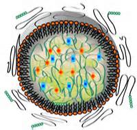
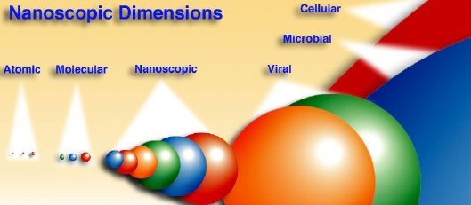
Above: Nanoparticles are larger than typical molecules but smaller than viruses. (They're labeled 'nanoscopic' in this diagram). They're similar in size to many proteins, which is part of the reason the can operate well inside of cells. Image courtesy University of Michigan-Ann Arbor.---Picture this: Before a space mission, an astronaut uses a hypodermic needle to inject a clear fluid, laced with nanoparticles, into his bloodstream. During flight, he puts a small device in his ear. This device, shaped like a hearing aide, uses a tiny laser to count glowing cells as they flow through capillaries in the eardrum. A wireless link relays those data to the spaceship's main computer for processing.--This sci-fi scenario is still at least 5 to 10 years away, but a lot of the necessary pieces are already taking shape in the laboratory[F4] .--That clear fluid injected into the astronaut's bloodstream would contain millions of microscopic nanoparticles. The nanoparticles themselves are nothing new: Scientists have been using them in the laboratory for at least 5 years, and they have employed them safely in lab animals.--The particular kind of nanoparticle that Baker uses resembles tumbleweed: a little ball-shaped bundle of branching "twigs" growing out from a central point. –

nanoparticles called dendrimers

By itself, this tumbleweed is inert. (That's good: it means it's not toxic.) It only serves as a generic platform upon which to build. All the useful functions of the nanoparticle -- seeking out the right kind of cells, detecting signs of radiation damage, offering up a fluorescent "red flag" -- come from molecules attached onto this scaffolding. The free ends of the twigs provide lots of binding points where these molecules can be attached (128 locations with the nanoparticles Baker's group uses)[F5] .

Right: The nanoparticles that Baker's group uses are called 'dendrimers,' and are built up by adding branching segments around a central core. Image courtesy University of Michigan-Ann Arbor. [More]--Choosing which molecules to attach is how scientists customize the nanoparticle to do their bidding. For example, Baker's group wants to tweak their nanoparticles to enter a kind of white blood cell called a lymphocyte, which is especially sensitive to radiation.--"How do we specifically target lymphocytes?" asks Thommey Thomas, a research assistant professor on Baker's team. "Because once you inject nanoparticles into the bloodstream they can go anywhere, right?"--"We had to find some specific targeting molecules on the surface of these lymphocytes," he explains.--All of the body's cells naturally have "receptor" molecules embedded on their outer surfaces. These receptors control which chemicals can enter the cell: for example, a kidney hormone in the bloodstream only enters kidney cells. By attaching a molecule to their nanoparticles that matches up with a specific receptor on lymphocytes, the researchers assure that these roaming nanoparticles wind up inside only the right cells[F6] .--Left: James Baker, director of the Center for Biologic Nanotechnology at the University of Michigan. [More]--Once inside the lymphocytes, nanoparticles need a way to detect radiation damage. One way is to watch for signs that the cell is about to self-destruct. Lymphocytes commit cellular suicide (called "apoptosis") when they've been damaged by radiation. This is a genetically programmed behavior carried out by special "suicide" enzymes. Baker's group has discovered how to attach to the nanoparticles a fluorescent dye molecule that reacts to these suicide enzymes. [F7] Lymphocytes beginning to self-destruct due to radiation damage would glow.--The research group has also developed a laser system to count the glowing cells. They've already shown that it can count cells in a mouse's bloodstream as those cells pass through the capillaries in its ear, but Baker says it's still too early to know what form this laser system would take for a space mission--maybe a micro-laser integrated into a hearing-aide-like device, he speculates.--The net result: continuous, real-time monitoring of radiation damage to the cells in an astronaut's bloodstream -- no bulky medical equipment required.

More Information

Center for Biologic Nanotechnology -- home page for the group at the University of Michigan performing the research discussed in this article

Voyage of the Nano-surgeons -- (Science@NASA) NASA-funded scientists are crafting microscopic vessels that can venture into the human body and repair problems -- one cell at a time.

Can People Go to Mars? -- (Science@NASA) Space radiation between Earth and Mars poses a hazard to astronauts. How dangerous is it out there? NASA scientists are working to find out.

Dr. James Baker -- biographical sketch for the director of the Center for Biologic Nanotechnology


*****************************************************************************************************************************************************


 Scientists resolve debate over how many bacteria fight off invaders

Date:May 7, 2015

Source:Rockefeller University

Every inch of our body, inside and out, is oozing with bacteria. In fact, the human body carries 10 times the number of bacterial cells as human cells. Many are our friends, helping us digest food and fight off infections, for instance. But much about these abundant organisms, upon which our life depends, remains mysterious. In research reported May 7 in Cell, scientists at Rockefeller finally crack the code of a fundamental process bacteria use to defend themselves against invaders.--For years, researchers have puzzled over conflicting results about the workings of a type of immune system found in many species of bacteria. Some data showed that, when a virus invaded a bacterial cell, this mechanism -- known as type III CRISPR-Cas -- would target the virus's DNA, preventing it from adopting the bacteria's machinery in order to copy itself and infect more bacteria. But other experiments suggested type III CRISPR-Cas could only disable a virus by cleaving the viral RNA.--Luciano Marraffini and Poulami Samai, both at Rockefeller, wanted to get to the bottom of this puzzle. In their experiments, Samai, a postdoctoral fellow, tested the cleavage of DNA and RNA by the type III CRISPR-Cas system. But she added a key ingredient no one else had before, a protein known as RNA polymerase, which the cell uses to transcribe DNA to RNA. She and Marraffini, head of the Laboratory of Bacteriology, saw that CRISPR-Cas did, indeed, cleave the RNA produced from a virus's DNA -- but it would also cleave the virus's DNA.--There are advantages to such a two-pronged system, says Marraffini. Many viruses integrate into the genomes of the cells they infect and remain dormant, he says, causing no harm. In fact, these viruses can be beneficial to bacteria, by carrying toxins that help bacteria promote their own survival, for instance. The diphtheria toxin, for instance, is secreted by a species of bacteria, but the gene encoding the toxin comes from a virus. "By requiring viruses to begin transcribing their DNA into RNA before disabling them, the type III CRISPR-Cas system leaves dormant viruses intact, allowing them to continue benefiting the bacteria that host them," he notes.-Learning the details of how microbes carry out their functions can have important implications for health and science, Marraffini says. Besides being an incredibly abundant form of life on the planet, fueling the health and disease of every species and ecosystem, microbes have been the source of a number of technological tools that have revolutionized science and medicine.--"More than forty years ago, scientists discovered enzymes that cut DNA from studying the viruses that infect bacteria, inspiring a new class of tools that created a revolution in biomedicine," says Marraffini. Now, new technology based on another type of CRISPR-Cas is leading another wave in that revolution, allowing scientists to quickly and easily manipulate genomes in ways they never could before. "This is a testament to how the basic biology of microbes can be very useful. Microbes are a crucial part of biology on the planet, and it's important to understand how they work."--Story Source-The above story is based on materials provided by Rockefeller University. -Journal Reference-Luciano Marraffini et al. Co-transcriptional DNA and RNA Cleavage during Type III CRISPR-Cas Immunity. Cell, May 2015 DOI: 10.1016/j.cell.2015.04.027 show

*************************************************************************

Researchers follow zinc to uncover pathway that fine-tunes brain signaling

A study team led by researchers at the University of Pittsburgh School of Medicine who used specially developed technologies to "follow the zinc" have uncovered a previously unknown pathway the brain uses to fine-tune neural signaling -- and that may play a role in Alzheimer's and other diseases. Their findings appear online this week in the Proceedings of the National Academy of Sciences.--Scientists have long observed the presence of bubble-like vesicles that contain the neurotransmitter glutamate and zinc at the synapses, specialized contacts among neurons where neurotransmitters are released to propagate electrical signals through the brain. Glutamate is the major excitatory neurotransmitter in the brain, but the need for synaptic zinc, an essential element that acts as a co-factor for many enzyme and regulatory proteins, has not been understood, said Thanos Tzounopoulos, Ph.D., associate professor in the Auditory Research Group, Department of Otolaryngology, Pitt School of Medicine.--"Until now, we haven't had the ability to quantify or follow zinc when it is released into the synaptic cleft," he said. "In this study, we employed new tools to do that and found a pathway that could be important for conditions such as Huntington's disease and Alzheimer's."Co-investigator Stephen Lippard, Ph.D., and his team at the Massachusetts Institute of Technology (MIT) developed an agent that fluoresces when it binds zinc, making it possible for the first time to measure zinc levels accurately and track the element's movements. They also created an agent that blocks zinc activity, thus allowing them to disrupt the metal's actions to determine its function.--The researchers learned that, indeed, zinc was released from vesicles and diffused from the release site. Surprisingly, it could bind to so-called extrasynaptic glutamate NMDA-type receptors, just like the neurotransmitter glutamate. Whereas glutamate activates these receptors, zinc inhibits them."Glutamate acts like an accelerator of neuronal activity, while zinc behaves like a brake that fine tunes that signal," Dr. Tzounopoulos said. "The receptors that zinc influences are thought to play a role in neurodegenerative diseases, so these findings could open new research avenues in the field."--Story Source-The above story is based on materials provided by University of Pittsburgh Schools of The Health Sciences. Note: Materials may be edited for content and length.-Journal Reference-Charles T. Anderson, Robert J. Radford, Melissa L. Zastrow, Daniel Y. Zhang, Ulf-Peter Apfel, Stephen J. Lippard, Thanos Tzounopoulos. Modulation of extrasynaptic NMDA receptors by synaptic and tonic zinc. Proceedings of the National Academy of Sciences, 2015; 201503348 DOI: 10.1073/pnas.1503348112 --University of Pittsburgh Schools of the Health Sciences. "Researchers follow zinc to uncover pathway that fine-tunes brain signaling." ScienceDaily. ScienceDaily, 4 May 2015. <www.sciencedaily.com/releases/2015/05/150504154950.htm>.

*******************************************************************

 Targeting cancer therapy with phosphoproteomics

Medulloblastomas (MB), the most common malignant pediatric brain tumor, originate from dysregulation of developmental signaling pathways. To discover important drug targets within these pathways, researchers have undertaken the first quantitative mass spectrometry-based phosphoproteomic approach to identify important phosphorylation events, using the Hh signaling pathway as the model.- Quantitative phosphoproteomic analysis was performed using SILAC (Stable Isotope Labeling with Amino acids in Cell culture), combined with strong cation exchange fractionation and phosphopeptide enrichment by immobilized metal affinity chromatography (IMAC), followed by multiplexed quantitative mass spectrometry- The study revealed changes in phosphorylation of 94 proteins only 25 minutes after Shh exposure. Motif analysis revealed a novel and critical role for the kinase, CK2, in mediating 45 percent of all early phosphorylation events. Importantly, CK2 affects terminal Hh signaling components, circumventing challenges of emergence of resistance and a priori resistance commonly encountered with existing small molecule inhibitors developed for medulloblastoma. CK2 inhibitors demonstrated early and sustained inhibition of Hh signaling across several mammalian cell types, including MB cells. In vivo, mice harboring flank MB allografts derived from Ptch+/−;Tpr53−/− tumor harbouring a point mutation in Smo that renders them resistant to other Hh pathway inhibitors, showed near-complete cessation of tumor growth in response to TBB, a highly potent and selective inhibitor of CK2.-- This quantitative phosphoproteomic approach to Hh signaling has provided a perspective that was unattainable with previous transcription and genome-based efforts. This success using one pathway will set the foundation for others to apply a similar approach in different tumor initiating pathways.--Story Source-The above story is based on materials provided by American Association of Neurological Surgeons (AANS). Note: Materials may be edited for content and length.--American Association of Neurological Surgeons (AANS). "Targeting cancer therapy with phosphoproteomics." ScienceDaily. ScienceDaily, 6 May 2015. <www.sciencedaily.com/releases/2015/05/150506182709.htm>. This quantitative phosphoproteomic approach to Hh signaling has provided a perspective that was unattainable with previous transcription and genome-based efforts. This success using one pathway will set the foundation for others to apply a similar approach in different tumor initiating pathways.--Story Source-The above story is based on materials provided by American Association of Neurological Surgeons (AANS). Note: Materials may be edited for content and length.--American Association of Neurological Surgeons (AANS). "Targeting

***********************************************************************

USDA Gives $3.8 Million in Grants to Develop and Promote Nanotech in Food


The US Department of Agriculture (USDA) is giving millions of dollars to universities across America for the development and study of nanotechnology to be used in food.--Despite research existing on the potential concerns over health and safety of nanotech in food, the USDA wants to push forward with nanotech in full force. This agency of the government is known for its revolving door relationship with Big Food.--A statement from the Center for Food Safety sums up the health concerns well--The subject of nanotechnology and our food supply offers an alarming view of the potential for human health issues. Amazingly, the U.S. government currently does not regulate the use of nanotechnology in food products, despite its widespread use and serious public health concerns. Europe and the Canadian government have taken the first steps to limit the use of nanotechnology in food, but the U.S. has so far only issued draft guidelines to companies.”--So what is this nanotechnology exactly? That’s a complicated question. According to the USDA, “Nanomaterials can occur naturally, for example in volcanic ash and ocean spray, and may also be incidental byproducts of human activity, such as homogenization or milling. They can also be produced intentionally with specific properties through certain chemical or physical processes.”--These intentionally created nanomaterials have been in use in the US food supply for over ten years — mostly in packaging and processing. According to UnderstandingNano.com,-“Clay nanocomposites are being used to provide an impermeable barrier to gasses such as oxygen or carbon dioxide in lightweight bottles, cartons and packaging films. Storage bins are being produced with silver nanoparticles embedded in the plastic. The silver nanoparticles kill bacteria[F1]  from any material that was previously stored in the bins, minimizing health risks from harmful bacteria.”--Nanotech is increasingly being pursued for use directly in the food we eat, rather than just in the packaging. According to Popular Mechanics:“The most commonly used nanoparticle in foods is titanium dioxide. It’s used to make foods such as yogurt and coconut flakes look as white as possible, provide opacity to other food colorings, and prevent ingredients from caking up. Nanotech isn’t just about aesthetics, however. The biggest potential use for this method involves improving the nutritional value of foods.-“Nano additives can enhance or prevent the absorption of certain nutrients. In an email interview with Popular Mechanics, Jonathan Brown, a research fellow at the University of Minnesota, says this method could be used to make mayonnaise less fattening by replacing fat molecules with water droplets.”-There has been a 1000% increase in nanotech used for food since 2008 and is now being deployed by major companies including Kraft, General Mills, Hershey, Nestle, Mars, Unilever, Smucker’s and Albertsons.-This is where the $3.8 million in USDA grants come in. According to the PDF released by the USDA, the grants entail; University of Wisconsin, Madison, WI $450,100 | Tailor polyanhydride nanoparticles to encapsulate and release antibiotics to protect shrimp against bacterial pathogens.”

“Pennsylvania State University, University Park, PA $447,788 | Obtain a basic understanding of starch-nanoclay interactions in dispersion; evaluate the disintegration, release, and antimicrobial properties of cross-linked, crystallized, and iodine-loaded starch fibers; determine the effect of alignment and drawing on thermomechanical properties  of starch fibers; and assess the feasibility of using a multi-jet electrospinning setup to scale the electrospinning process for starch fiber production.”

“Rutgers University, New Brunswick, NJ $450,000 | Complete a national survey that will examine the acceptance of food nanotechnology; assess consumers’ beliefs about the relationship of nanotechnology to healthfulness; evaluate acceptability of nanomaterials in functional foods and pet food applications; examine the acceptable characteristics of nano-enabled smart food packaging; assess use value of visuals communicating the potential for nanotechnology; and examine how consumers use visuals to interpret nanotechnology concepts.”

The Rutgers University grant reveals that the USDA will be funding market research to directly benefit the businesses seeking to manufacture and market nanotechnology for food.-What are the real implications of this? There’s one thing for sure; the element of power is a very necessary thing to consider. With all technology like this, monopolization and hierarchical structures are a potential problem. The USDA and other health oversight agencies have a long track record of approving controversial practices used in our food that later turn out to have deadly health and environmental impacts. Understanding the current players in the agro-tech business including Monsanto and Syngenta, you can see how this could end badly.-The implications of this are significant for the future of Agorism, sustainable food, independent agricultural business, monopolization of food, the health of the people consuming this food, and much more.-No one is saying that nanotech in food is inherently bad, but with the history of the organizations funding this technology, people are naturally suspicious. More research is needed and concerns must be addressed before nanotech in our food becomes our next big mistake.-Please share this with as many people as possible. It is relevant to everyone.-This article is free and open source. You have permission to republish this article under a Creative Commons license with attribution to the author and TheAntiMedia.org. Tune in! The Anti-Media radio show airs Monday through Friday @ 11pm Eastern/8pm Pacific. Image credit: National Cancer Institute. Help us fix our typos: edits@theantimedia.org.


*****************************************************************************

Soft robot- Shaping itself and moving with own internally generated power

Date: May 5, 2015

Source: University of Pittsburgh

Summary:What if a new material would allow for development of a 'soft robot' that could reconfigure its own shape and move using its own internally generated power?


Time evolution of a rectangular SP-BZ gel in non-uniform light; time increases from (a) to (d). Both ends of the sample are exposed to light; the central circular region is not illuminated.

For decades, robots have advanced the efficiency of human activity. Typically, however, robots are formed from bulky, stiff materials and require connections to external power sources; these features limit their dexterity and mobility. But what if a new material would allow for development of a "soft robot" that could reconfigure its own shape and move using its own internally generated power?--By developing a new computational model, researchers at the University of Pittsburgh's Swanson School of Engineering have designed a synthetic polymer gel that can utilize internally generated chemical energy to undergo shape-shifting and self-sustained propulsion. Their research was published April 30th in the journal Scientific Reports, published by Nature.--The authors are Anna C. Balazs, PhD, the Swanson School's Distinguished Professor of Chemical and Petroleum Engineering and the Robert v. d. Luft Professor; and Olga Kuksenok, PhD, --Research Associate Professor.--"Movement is a fundamental biological behavior, exhibited by the simplest cell to human beings. It allows organisms to forage for food or flee from predators. But synthetic materials typically don't have the capability for spontaneous mechanical action or the ability to store and use their own energy, factors that enable directed motion" Dr. Balazs said. "Moreover in biology, directed movement involves some form of shape changes, such as the expansion and contraction of muscles. So we asked whether we could mimic these basic interconnected functions in a synthetic system so that it could simultaneously change its shape and move."-As a simple example in nature, Drs. Balazs and Kuksenok use the single-celled organism euglena mutabilis, which processes energy to expand and contract its shape in order to move. To mimic the euglena's mobility, Drs. Balazs and Kuksenok looked to polymer gels containing spirobenzopyran (SP) since these materials can be morphed into different shapes with the use of light, and to Belousov-Zhabotinsky (BZ) gels, a material first fabricated in the late 1990s that not only undergoes periodic pulsations, but also can be driven to move in the presence of light.-"The BZ gel encompasses an internalized chemical reaction so that when you supply reagents, this gel can undergo self-sustained motion," Dr. Kuksenok explains. "Although researchers have previously created polymer chains with both the SP and BZ functionality, this is the first time they were combined to explore the ability of "SP-BZ" gels to change shape and move in response to light."-As Balazs and Kuksenok noted, these systems are distinctive because they not only undergo self-bending or folding, but also self-propelled motion. Namely, the material integrates the powerful attributes of each of the components-the ability of SP-functionalized gels to be "molded" with light and the autonomous mechanical actions of the BZ gels.-According to Dr. Balazs, there were unexpected results during their research. "Uniform light exposure won't work. We had to place the light at the right place in order for the gel to move. And if we change the pattern of the light, the gel displays a tumbling motion.-"We also found that if we placed the SP in certain regions of the BZ gel and exposed this material to light, we could create new types of self-folding behavior." The next phase of the research will be to combine the patterning of the SP and BZ functionality in the gels with the patterning of the light to expand the polymer's repertoire of motion.--Dr. Balazs adds that these SP-BZ gels could enable the creation of small-scale soft robotics for microfluidic devices that can help carry out multi-stage chemical reactions.--"Scientists are interested in designing biomimetic systems that are dissipative -- they use energy to perform a function, much like our metabolism allows us to carry out different functions," she explained. "The next push in materials science is to mimic these internal metabolic processes in synthetic materials, and thereby, create human-made materials that take in energy, transform this energy and autonomously perform work, just as in biological systems."--The benefit of using polymer gels instead of metals and alloys to build a robot is that it greatly reduces its mass, improves its potential range of motion and allows for a more "graceful" device.--"To put it simply, in order for a robot to be able to move more autonomously in a more biomimetic way, it's better if it's soft and squishy," Dr. Kuksenok says. "It's ability to grab and carry something isn't impeded by non-flexible, hard edges. You'd also like its energy source incorporated into the design so that it's not carrying that as extra baggage. The SP-BZ gel is pointing us in that direction."--Story Source-The above story is based on materials provided by University of Pittsburgh. Note: Materials may be edited for content and length.-Journal Reference-Olga Kuksenok, Anna C. Balazs. Designing Dual-functionalized Gels for Self-reconfiguration and Autonomous Motion. Scientific Reports, 2015; 5: 9569 DOI: 10.1038/srep09569

***********************************************************************


TOP A


 [F1]Including yours---brain damage—liver damage—skeletal damage-spleen damage and the oxidation of healthy cells throughout the body and the use with nanobots inside the body

 [F2]NanoParticles—are a billionth in size—and with the intro on nano becoming incorporated in everything today the discarding and waste management of these particulates are becoming more prevalent

 [F3]They are talking micron levels which is a millionth in size

 [F4]In other words they had the social network they had in there homeland and that was transferred with them---so environmental factors then would have been minimized stress wise due to the network of the cultural connection---which the ones who have been assimilated would not have that type of support

And immigrants usually will stick together even if they are from differing countries—due to the social aspect

 [F5]Studies done on what could get effected in the Body

 [F6]So this will imply strongly we will need something more effective to reduce or remove these out of the body

 [F7]Best to avoid any and all products made with any nano compostions—since there is no way to prevent the harmful effects—that  so far can be determined

 [F8]Interesting ---a explosion  and the reactor is in safe stable –hmm I am reall y impressed with this fantasy

 [F9]I am again impressed

 [F10]Seems to me it was not exactly ready---potential sabotage??just a thought

*********************************************************************

TOP B

HOME


Show of the Month May 16 2015


Link between vitamin E, exposure to air pollution


Saffron and retina- neuroprotection and pharmacokinetics

Study shows where damaged DNA goes for repair

Scientists Find Alarming Deterioration In DNA Of The Urban Poor

Is the toxic potential of nanosilver dependent on its size?

Nanoparticles can damage DNA, increase cancer risk

Explosion rocks nuclear power plant in New York


*********************************************************************

Link between vitamin E, exposure to air pollution

­ A new study from King's College London and the University of Nottingham has found an association between the amount of vitamin E in the body, exposure to particulate pollution and lung function. The paper adds to growing evidence from previous studies suggesting that some vitamins may play a role in helping to protect the lungs from air pollution. Although the new study did not specifically demonstrate a protective effect, it is the first to show a clear link between vitamin E concentrations in the blood and exposure to fine particulate pollution in the general population.-- Particulate matter (PM) [F2] is one of the main air pollutants thought to be damaging to human health. Previous studies have reported an inverse association between exposure to PM and lung function. However, the underlying mechanisms linking ambient air pollution to lung function are not yet fully understood--The new data, published in the American Journal of Respiratory and Critical Care Medicine, looked at the association between lung function and a set of metabolites -- chemical signatures circulating in the blood -- and between these metabolites and exposure to PM10 and PM2.5 (particles smaller than 10 and 2.5 microns, respectively)[F3]  determined as the concentrations of these pollutants at the participants' residence-Two-hundred and eighty metabolites were measured in the blood of over 5,500 fasting volunteers from the Twins UK study who had also undergone a spirometry or lung test. This test determines the lung's forced vital capacity (FVC), a measure of the amount of air you can exhale with force after you inhale as deeply as possible, and forced expiratory volume (FEV), a measure of the amount of air you can exhale with force in one breath--. A subset of this group of twins -- around 500 participants -- living in the Greater London area also had their long-term exposure to PM estimated from their postcode using computer modelling of air pollution across London. Participants completed a medical history and lifestyle questionnaire, including questions on whether they took vitamin supplements--  The profiling revealed 13 metabolites significantly associated with FVC, 10 of which were also identified for FEV. Of the metabolites associated with lung function, eight were also significantly associated with exposure to both PM2.5 and PM10. In all eight instances, a higher exposure to PM was found to correlate with lower levels of the metabolite and a lower FEV.-- Among the eight metabolites identified were two well-known antioxidants, alpha tocopherol or a-tocopherol (biologically active form of Vitamin E) and a metabolite of ascorbic acid (Vitamin C) known as threonate. Both compounds have previously been linked to lung function as well as exposure to PM-The strongest association both with PM2.5 and FEV was seen with vitamin E. Individuals with a higher exposure to PM2.5 had significantly lower levels of alpha-tocopherol and also had lower lung function. These findings provide further evidence supporting the theory that PM damages lungs through oxidative attack while alpha-tocopherol acts to minimise oxidative injury.-Dr Ana Valdes, Reader at the University of Nottingham and co-author of the study, explained: 'Our work builds on a number studies exploring whether some vitamins can counteract the negative effect on lungs caused by air pollution. More work is needed to establish whether antioxidant supplements do indeed provide protection to the lungs in the general population.'-Professor Frank Kelly, Head of the Environmental Research Group at King's College London and co-author of the study, said: 'These new findings are consistent with previous reports which observed lower levels of vitamin E in people with lung conditions such as asthma. However, we do not yet fully understand which types of particulate pollution specifically damage the lungs or which vitamins best interfere with this pathway to reduce the level of damage-- Story Source--The above story is based on materials provided by King's College London.

****************************************************************************

Saffron and retina- neuroprotection and pharmacokinetics.

Vis Neurosci. 2014 Sep;31(4-5):355-61

Authors: Bisti S, Maccarone R, Falsini B

Abstract
Age-related macular degeneration (
AMD) is a retinal neurodegenerative disease whose development and progression are the results of a complex interaction between genetic and environmental risk factors. Both oxidative stress and chronic inflammation play a significant role in the pathogenesis of AMD. Experimental studies in rats with light-induced photoreceptors degeneration demonstrated that saffron may protect photoreceptor from retinal stress, preserving both morphology and function and probably acting as a regulator of programmed cell death, in addition to its antioxidant and anti-inflammatory properties. Recently, a randomized clinical trial showed that in patients with early AMD, dietary supplementation with saffron was able to improve significantly the retinal flicker sensitivity suggesting neuroprotective effect of the compound. Here, we examine the progress of saffron dietary supplementation both in animal model and AMD patients, and discuss the potential and safety for using dietary saffron to treat retinal degeneration. --PMID: 24819927 [PubMed - indexed for MEDLINE]

**********************************************************************

Study shows where damaged DNA goes for repair

Date:May 3, 2015

Source:Tufts University


Summary-New research sheds light on the process of DNA repair in the cell. Expanded repeats of the CAG/CTG trinucleotide in yeast shift to the periphery of the cell nucleus for repair. This shift is important for preventing repeat instability and genetic disease. Going out to the 'repair shop' at the nuclear periphery is a previously unrecognized yet important step to maintain repetitive DNA and to prevent damage to chromosomes, researchers report--A Tufts University study sheds new light on the process by which DNA repair occurs within the cell. In research published in the May 15 edition of the journal Genes & Development and available May 4 online in advance of print, Tufts University biologist Catherine Freudenreich and her co-authors show that expanded repeats of the CAG/CTG trinucleotide (CAG) in yeast shift to the periphery of the cell nucleus for repair. This shift is important for preventing repeat instability and genetic disease.--CAG expansions are significant because they are at the root of several neurodegenerative and neuromuscular diseases such as Huntington's disease, myotonic dystrophy and multiple subtypes of spinal cerebella ataxia.--Short triplet repeats do not always cause problems. However, sometimes short triplet repeats expand and become longer than normal. The expanded repeat sequences change the shape of the DNA molecule from a double helix into a hairpin-like structure that is difficult for the cellular machinery to replicate and repair and can cause breakage of the DNA molecule.--Disease occurs when the numbers of expanded CAG trinucleotide repeats exceed a stability threshold. For Huntington's disease, the threshold is 38 to 40 repeats. Myotonic dystrophy results when there are close to 200 repeats.--"Appropriate repair of DNA damage at CAG repeats is vital to the cell as proper repair can prevent further expansion and aggravation of the disease," says Freudenreich.--In their study, "Regulation of Recombination at Yeast Nuclear Pores Controls Repair and Triplet Repeat Stability," the researchers introduced CAG repeats of different sizes into budding yeast chromosomes. The repeat-containing chromosomes were tagged with a fluorescent molecule, so that their location in the cell nucleus could be followed visually.--"We found that the chromosomes containing the expanded CAG repeats moved from the interior of the nucleus to the nuclear periphery to be repaired," says Freudenreich. "Our research shows that the DNA with expanded repeats makes this 'trip' for repairs. Proximity to the nuclear envelope helps the repair of the repeat to occur without mistakes."-"When the movement of the DNA molecule was prevented, the chromosomes broke more frequently, and the repeat was more likely to be mutated," says Freudenreich. "Thus, going out to the 'repair shop' at the nuclear periphery is a previously unrecognized yet important step for repetitive DNA to be maintained and to prevent damage to chromosomes."--"The longer the repeat, the greater the frequency of relocation," Freudenreich notes. "For example, CAG-130 relocates more often than CAG-70, and an unexpanded CAG-15 does not relocate more than a non-repeat control. We think if the replication machinery stalls at the repeat it triggers relocation."--Normally, each single strand of DNA serves as a template for remaking the other strand. The enzymes involved in DNA replication rebuild each strand to make two chromosomes out of one. One section of the double-stranded DNA molecule is separated into two single strands. The resulting Y-shaped structure is called the replication fork. This process stalls when there is a problem with the DNA.--At the periphery, the damaged DNA interacts with the nuclear pore complexes (NPCs), a complex of proteins that serves as gatekeeper between the cell's nucleus and the surrounding cytoplasm. One of these gatekeepers is the Nup84 complex and the associated Slx5/8 complex. They are present at every NPC.--The research team showed that both of these complexes are needed for repeat DNA to be repaired properly. The Slx5/8 complex plays a vital role: it is required for tethering the repeat DNA to the NPC, and it appears to regulate a known repair protein (Rad52) in order to facilitate appropriate repair, thereby preventing repeat expansions and chromosome breakage.--"The nuclear pore complex plays a role in preventing chromosome breaks as well as preventing instability of the CAG repeat, so it has a role in DNA repair," says Freudenreich. Story Source-The above story is based on materials provided by Tufts University. Note: Materials may be edited for content and length. Journal Reference-Xiaofeng A. Su, Vincent Dion, Susan M. Gasser, Catherine H. Freudenreich. Regulation of recombination at yeast nuclear pores controls repair and triplet repeat stability. Genes & Development, 2015; DOI: 10.1101/gad.256404.114


************************************************************************

Scientists Find Alarming Deterioration In DNA Of The Urban Poor

The urban poor in the United States are experiencing accelerated aging at the cellular level, and chronic stress linked both to income level and racial-ethnic identity is driving this physiological deterioration.--These are among the findings published this week by a group of prominent biologists and social researchers, including a Nobel laureate. Dr. Arline Geronimus, a visiting scholar at the Stanford Center for Advanced Study and the lead author of the study, described it as the most rigorous research of its kind examining how "structurally rooted social processes work through biological mechanisms to impact health."

What They Found

Researchers analyzed telomeres of poor and lower middle-class black, white, and Mexican residents of Detroit. Telomeres are tiny caps at the ends of DNA strands, akin to the plastic caps at the end of shoelaces, that protect cells from aging prematurely. Telomeres naturally shorten as people age. But various types of intense chronic stress are believed to cause telomeres to shorten, and short telomeres are associated with an array of serious ailments including cancer, diabetes, and heart disease. Evidence increasingly points to telomere length being highly predictive of healthy life expectancy. Put simply, "the shorter your telomeres, the greater your chance of dying."-The new study found that low-income residents of Detroit, regardless of race, have significantly shorter telomeres than the national average. "There are effects of living in high-poverty, racially segregated neighborhoods -- the life experiences people have, the physical exposures, a whole range of things -- that are just not good for your health," Geronimus said in an interview with The Huffington Post.--But within this group of Detroit residents, the ways in which race-ethnicity and income were associated with telomere length were strikingly varied. -White Detroit residents who were lower-middle-class had the longest telomeres in the study. But the shortest telomeres belonged to poor whites. Black residents had about the same telomere lengths regardless of whether they were poor or lower-middle-class. And poor Mexicans actually had longer telomeres than Mexicans with higher incomes.-Geronimus said these findings demonstrated the limitations of standard measures -- like race, income and education level -- typically used to examine health disparities. "We've relied on them too much to be the signifiers of everything that varies in the life experiences of difference racial or ethnic groups in different geographic locations and circumstances," she said. --According to Geronimus, it's important to consider not just racial-ethnic identity, but also "the extent to which it is validated, or discriminated against, or even understood within your everyday life experience" can affect an individual's health dramatically, Geronimus said. "Race is not race is not race. Poverty is not poverty is not poverty. Early health deterioration is sensitive to a broad range of life experiences."

When Ethnic Identity Impacts Health

So why did poor Mexicans in this study have longer (i.e., generally healthier) telomeres than the nonpoor Mexicans? Geronimus first noted that most poor Mexicans in Detroit were either first-generation immigrants to the United States or part of close-knit ethnic enclaves[F4] . In contrast, nonpoor Mexicans were more often born in the U.S. and were more integrated into American culture through work or school.--"If they're immigrants, then they come with a different cultural background and upbringing that didn't stress that as Mexicans they were somehow 'other' or 'lesser' than other Americans," said Geronimus. "They come with a set of support systems and with a cultural orientation that doesn't undermine their sense of self-worth. They then often live in these ethnic enclaves, many of them don't speak anything other than Spanish, and so they're not interacting with Americans who view them as 'other' or who treat them badly. It's not that they're immune to that treatment but they're not as sensitive to it and they also just don't experience it as often."--On the other hand, nonpoor Mexicans are more likely to be "exposed to some of the negative views of Mexicans held by some Americans, the conflation of anyone of Mexican origin as being an immigrant or possibly an undocumented immigrant, or even more neutral assumptions like 'they must speak Spanish,' or 'they don't understand English.'" Ironically, in seeking to become socially mobile and avoid the stress of poverty, these lower-middle-class Mexicans may face even more pronounced stressors tied to their ethnic identity.--Geronimus said the findings of the new study, based on quantitative physiological research, "line up perfectly" with previous ethnographic studies of Mexicans in Detroit done by another researcher, Dr. Edna Viruell-Fuentes of the University of Illinois at Urbana-Champaign. A health site summarized the conclusions of Viruell-Fuentes's work--In 2007, Viruell-Fuentes interviewed 40 first- and second-generation Mexican immigrant women in the Detroit area. Though she points out that racial dynamics are hard to measure, based on her interviews Viruell-Fuentes believes that "identifying experiences that are discriminatory may be a learned process." Often first-generation women characterized certain interactions as simply rude, while second-generation women labeled similar experiences as discriminatory. [...]--In a 2012 review paper, Viruell-Fuentes pointed out that the first generation tends to stay within ethnic enclaves that may buffer some of the social disadvantages that immigrants face. "For the second generation, what I think is different, is that they have a lifelong exposure to an environment that stigmatizes their identity, which in turn can affect their health negatively," she said.--"Often the question is raised, what is it about immigrants that makes them more resilient?" Viruell-Fuentes said. "But the other piece of the question for me is, what is it about the United States that is damaging to people's health?--Other health effects tied to race-ethnicity identified in the new study could be viewed as counterintuitive. Income level seemed to have no effect on the telomere lengths of black Detroit residents, while the telomeres of poor whites were significantly shorter than those of nonpoor whites. Why? -The study's authors noted---Much research suggests the separation between poor and nonpoor blacks in everyday life is less marked than between poor and nonpoor whites. Not only do blacks tend to have greater residential proximity owing to residential segregation, but often poor and the nonpoor blacks are members of the same families and social networks, practice reciprocal obligations, or have similar experiences of cycling between low and moderate incomes. --Income instability among middle-class blacks reflects job insecurity, a relative lack of conventional assets or wealth to tide them over in rough times, or a network-level division of labor whereby some are expected to contribute to family economies through income generating work, others contribute by seeing to the family caretaking needs that facilitate the employment of others, and still others provide important services and skills as barter exchange.--Researchers also highlighted the hypersegregation in the Detroit area. "Most blacks in our sample live almost exclusively with other blacks (97% of Eastside Detroit residents are black) or are the majority group in integrated neighborhoods (e.g., 70% of Northwest Detroit residents are black), [and] whites are a clear minority in all of our Detroit areas (ranging from 2% to 21% of residents)."--They found that associations between telomere length and perceptions of neighborhood physical environment and neighborhood satisfaction were strongest for blacks, and questioned whether "safety stress, physical environment, and neighborhood satisfaction tap into a more global construct of how black participants experience Detroit neighborhoods, which on balance may be more positively than for white or Mexican participants."--In contrast, regarding white Detroit residents, the researchers wrote, "Perhaps with the exodus of most whites and many jobs from Detroit, the shrinking benefits of labor union membership and public pensions, and the overall reduction in taxation-based city services, the poor whites who remain are particularly adversely affected by the social and ecological consequences of austerity urbanism. Lacking the financial resources, social networks, and identity affirmation of the past, remaining Detroit whites may have less to protect them from the health effects of poverty, stigma, anxiety, or hopelessness in this setting."--Geronimus summarized, "I think a lot of people just don’t understand how bad it is for some Americans. It’s disproportionately people of color given our history of residential segregation and racism, but it’s also anyone who gets caught. It’s like the dolphins who get caught in the fishing nets, it’s anyone who gets caught there. If anything, some of our evidence suggests that whether it’s the poor Mexican immigrant or the African-Americans who have been discriminated against and dealt with hardship for generation after generation, they’ve developed systems to cope somewhat that perhaps white Detroiters haven’t. So there’s great strength in these populations. But it's not enough to solve these problems without the help of policymakers and more emphatic fellow citizens."

Telomeres, Health, And Social Justice

One co-author of this new study is Dr. Elizabeth Blackburn. She helped to discover telomeres, an achievement that won her the Nobel Prize in physiology in 2009.

When her research began in the mid-1970s, Blackburn worked on identifying telomeres in one-celled organisms she laughingly calls "pond scum." But over the years, as she and other scientists discovered the far-reaching human health implications of telomeres, her focus shifted.--"So much of what makes people either well-being or not is not coming from within themselves, it's coming from their circumstances. It makes me think much more about social justice and the bigger issues that go beyond individuals," she said in an interview from her office at the University of California San Francisco.--Blackburn believes that vital questions relevant to social policy have remained unanswered because the issues were highly complex and it was easy to question data from qualitative research methods, like people's questionnaire answers about their personal experiences and perceptions. "When something's really hard to assess, the easy thing is to dismiss it. They say it's soft science, it's not really hard-based science."--But now telomere data is providing a new way to quantitatively analyze some of these complex topics. Blackburn ticked off a list of studies in which people's experiences and perceptions directly correlated with their telomere lengths: whether people say they feel stressed or pessimistic; whether they feel racial discrimination towards others or feel discriminated against; whether they have experienced severely negative experiences in childhood, and so on. --"These are all really adding up in this quantitative way," she said. "Once you get a quantitative relationship, then this is science, right?"

**************************************************************************

Is the toxic potential of nanosilver dependent on its size?

Anna Huk12, Emilia Izak-Nau23, Bogumila Reidy4, Matthew Boyles2, Albert Duschl2, Iseult Lynch5 and Maria Dušinska1*

·         * Corresponding author: Maria Dušinska maria.dusinska@nilu.no

Author Affiliations

Particle and Fibre Toxicology 2014, 11:65  doi:10.1186/s12989-014-0065-1

Published: 3 December 2014

Abstract

Background

Nanosilver is one of the most commonly used engineered nanomaterials (ENMs). In our study we focused on assessing the size-dependence of the toxicity of nanosilver (Ag ENMs), utilising materials of three sizes (50, 80 and 200 nm) synthesized by the same method, with the same chemical composition, charge and coating.

Methods

Uptake and localisation (by Transmission Electron Microscopy), cell proliferation [F5] (Relative growth activity) and cytotoxic effects (Plating efficiency), inflammatory response (induction of IL-8 and MCP-1 by Enzyme linked immune sorbent assay), DNA damage (strand breaks and oxidised DNA lesions by the Comet assay) were all assessed in human lung carcinoma epithelial cells (A549), and the mutagenic potential of ENMs (Mammalian hprt gene mutation test) was assessed in V79-4 cells as per the OECD protocol. Detailed physico-chemical characterization of the ENMs was performed in water and in biological media as a prerequisite to assessment of their impacts on cells. To study the relationship between the surface area of the ENMs and the number of ENMs with the biological response observed, Ag ENMs concentrations were recalculated from μg/cm2 to ENMs cm2/cm2 and ENMs/cm2.

Results

Studied Ag ENMs are cytotoxic and cytostatic, and induced strand breaks, DNA oxidation, inflammation and gene mutations. Results expressed in mass unit [μg/cm2] suggested that the toxicity of Ag ENMs is size dependent with 50 nm being most toxic. However, re-calculation of Ag ENMs concentrations from mass unit to surface area and number of ENMs per cm2 highlighted that 200 nm Ag ENMs, are the most toxic. Results from hprt gene mutation assay showed that Ag ENMs 200 nm are the most mutagenic irrespective of the concentration unit expressed.

Conclusion

We found that the toxicity of Ag ENMs is not always size dependent. Strong cytotoxic and genotoxic effects were observed in cells exposed to Ag ENMs 50 nm, but Ag ENMs 200 nm had the most mutagenic potential. Additionally, we showed that expression of concentrations of ENMs in mass units is not representative. Number of ENMs or surface area of ENMs (per cm2) seem more precise units with which to compare the toxicity of different ENMs.


***************************************************************************

Nanoparticles can damage DNA, increase cancer risk

April 17th, 2007 in Nanotechnology / Bio & Medicine

Tissue studies indicate that nanoparticles, engineered materials about a billionth of a meter in size, could damage DNA and lead to cancer, according to research presented at the 2007 Annual Meeting of the American Association for Cancer Research.-- Nanoparticles are small enough to penetrate cell membranes and defenses, yet they are large enough to cause trouble by interfering with normal cell processes, researchers at the University of Massachusetts say. --Such nanoparticles are currently in use in electronics, cosmetics, and chemical manufacturing, among others industries. Because of their extremely small size, they can be difficult to isolate from the larger environment, as they are much too small for removal by conventional filtering techniques[F6] .--When nanoparticles find their way into cancer cells, they can wreak havoc, according to Sara Pacheco, an undergraduate researcher at the University of Massachusetts. Yet very little is known about how they behave in the environment or how they interact with and affect humans-"Unfortunately, only a very small portion of research on nanoparticles is focused on health and safety risks, or on threats to the environment," Pacheco said. "I am concerned because so many new nanoparticles are being developed and there is little regulation on their manufacture, use and disposal."- Pacheco and her colleagues looked at how two different types of nanoparticles could cause DNA damage in the MCF-7 line of breast cancer cells-- She and her team examined the genotoxicity of silica and C60 fullerene nanoparticle suspensions using the alkaline single-cell gel electrophoresis assay (Comet assay) to quantify breaks in single and double stranded DNA. The team chose these particular nanoparticle types because they are commonly used commercially – in electronics, textiles and sporting goods – and easy to work with in the laboratory setting.-- "We observed both dose-dependent and time-dependent increases in DNA damage in breast cancer cells exposed to either aqueous colloidal silica or C60 fullerenes," Pacheco said. "The DNA damage could potentially lead to mutations and ultimately increase the risk of cancer- One problem is that, while it’s clear that some nanoparticles can be more toxic than others, there’s not enough data as yet to determine the most dangerous types "A lot is unknown about nanoparticle function, but clearly both size and composition are important," Pacheco said. "Several studies have shown that smaller particles are more likely to enter cells and cause more toxicity-According to Pacheco, what makes matters worse is the fact that so far, aside from preventing their release, there are no known ways to prevent the harmful effects of environmental nanoparticles[F7] .-"It is important to know whether the nanoparticles are entering the cell and causing DNA damage directly or if they are acting on the membrane and inducing a cascade of events resulting in DNA damage," Pacheco said. "Once we understand the mechanisms by which nanoparticles induce their toxicity, we will be better able to prevent or mitigate their harmful effects."--In the meantime, the experimental team suggests that great caution should be taken in handling such nanoparticle suspensions and that any uncontrolled release should be avoided-Until we understand which types of nanoparticles are harmless and which have the potential to be harmful, I think it is prudent to limit their introduction into the environment," recommended Pacheco.--Source: American Association for Cancer Research

*****************************************************************************

Explosion rocks nuclear power plant in New York

An explosion has rocked a nuclear facility in the US state of New York, triggering fears across the country, reports say.

The explosion happened on Saturday at the Indian Point nuclear plant in the upstate part of New York.--According to the company running the plant, Entergy Corporation, the affected parts of the facility were "safely shut down" and in "safe and stable condition.”--There was no damage to the reactor[F8] , the most important part of a nuclear facility, according to US media reports. Authorities at the scene reported no human casualties either[F9] . --“The plant’s unit 3 reactor was closed after the explosion but the other one, Unit 2 is still operating,” Entergy Corporation spokesman Jerry Nappi told reporters.--“No threat to public safety at any time,” said the company, attempting to comfort the public as many expressed fears across the state.--People who were nearby the nuclear plant and witnessed the explosion said that there was a large blast followed by fire and then smoke.--The same nuclear plant was just brought back online on Friday after it was shut down for technical repairs[F10] .--The plant was built in 1962, but the current reactors operating went online in the late 70s.--The Indian Point nuclear center produces 25 percent of New York City’s and many suburbs across the state.--Over 1,600 employees work there and it produces over 2,000 megawatts of energy annually

TOP B


 [F1]Don’t you love the sales pitch here –incredible ---actually putting spyware in a human ---and utilizing this as a

health aide to sell this concept---when anyone who is anyone knows that nano does not opeate the way normal cells do and with the inclusion of nano in the body would overwrite the genetic codes---it would insert it’s artificial intelligence into a person and take over

 [F2]Am I hearing

bioweapon

 [F3]This is all meaningless---you add a nano program into the person and what it is going to show is the rate of exposure and rate of decay –if there is not the equipment to repair then what would be the point

 [F4]The laboratory would be the general public—morgellons is a form of nano poisoning---they have released this to see how the bugs can e taken out

Before they  perfect this for military use

 [F5]This is how the nano particles work on us as well

 [F6]This sounds good in theory But the reality is ---not accurate---and what happens once they are in those organs or tissue?? And hw does one extract them out?

 [F7]Some have seen these dyes on the skin they glow

********************************************************************************************************************************************

TOP C


Show of the Month May 23 2015


Pooled analysis confirms vitamin E as a treatment for non-alcoholic steatohepatitis


How vitamin E keeps muscles healthy


Obesegons


Contaminants that cause Obesegons


Atrazine –Endocrine Damaging –and Disorienteering Sexual Development


**************************************************************************

Pooled analysis confirms vitamin E as a treatment for non-alcoholic steatohepatitis

Date:April 23, 2015

Source:European Association for the Study of the Liver

Summary:

Vitamin E (d-alpha-tocopherol) is an effective treatment for non-alcoholic steatohepatitis[F1]  (NASH), according to new research. NASH occurs when the liver becomes inflamed due to the accumulation of fat. Over time, persistent inflammation can lead to the formation of fibrous scar tissue in the liver and around its blood vessels, which can eventually cause cirrhosis.


CITE Page

European Association for the Study of the Liver. "Pooled analysis confirms vitamin E as a treatment for non-alcoholic steatohepatitis." ScienceDaily. ScienceDaily, 23 April 2015. <www.sciencedaily.com/releases/2015/04/150423085348.htm>

Results revealed at The International Liver Congress™ 2015 show that vitamin E (d-alpha-tocopherol) is an effective treatment for non-alcoholic steatohepatitis (NASH). NASH occurs when the liver becomes inflamed due to the accumulation of fat. Over time, persistent inflammation can lead to the formation of fibrous scar tissue in the liver and around its blood vessels, which can eventually cause cirrhosis.--- A pooled analysis of data from two randomised trials comparing vitamin E versus placebo, and the placebo group from another trial comparing vitamin E use versus non-use, demonstrates that the efficacy of vitamin E is comparable to other treatments for NASH, including pioglitazone, metformin and obeticholic acid. In addition, treatment with vitamin E is associated with significant improvements in both NASH histology (45% vs 22% in those not treated with vitamin E) and resolution of disease (38% vs 20% in those not treated with vitamin E). There was no increase in cardiovascular events and no adverse lipid profiles were observed with vitamin E treatment-- A total of 347 patients (155 treated with vitamin E, 192 not treated with vitamin E) were included in the analysis which compared data from three clinical trials that investigated the efficacy and safety of vitamin E as a treatment for NASH: the PIVENS, TONIC and FLINT trials. Histologic improvement was defined as ? 2 point improvement in NAS with no worsening of fibrosis, and NASH resolution measured effectiveness.-- The study supports the use of vitamin E as a treatment for NASH-Story Source--The above story is based on materials provided by European Association for the Study of the Liver. Note: Materials may be edited for content and length

*************************************************************************


How vitamin E keeps muscles healthy

Date:

May 19, 2015

Source:

Medical College of Georgia at Georgia Regents University

Summary:

Body builders have it right: Vitamin E does help build strong muscles, and scientists appear to have figured out one important way it does it. Vitamin E has long known as a powerful antioxidant, and now scientists have shown that without it, the plasma membrane, which essentially keeps a cell from spilling its contents and controls what moves in and out, cannot properly heal.


Body builders have it right: vitamin E does help build strong muscles, and scientists appear to have figured out one important way it does it.

Vitamin E has long known as a powerful antioxidant, and now scientists have shown that without it, the plasma membrane, which essentially keeps a cell from spilling its contents and controls what moves in and out, cannot properly heal.

That's a big problem for many cells, such as muscle cells, which get membrane tears just from being used.-"Every cell in your body has a plasma membrane, and every membrane can be torn," said Dr. Paul L. McNeil, cell biologist at the Medical College of Georgia at Georgia Regents University and corresponding author of the study in the journal Free Radical Biology and Medicine.-The scientist suspects knowing the cell membrane repair action of vitamin E has implications for muscular dystrophy, and common diabetes-related muscle weakness, as well as traumatic brain injury, resulting from collisions on a football field, battlefield, or roadway. With a traumatic brain injury, for example, one of the first events that happens is that the plasma membrane of the neurons, key cells in the central nervous system, tear. "Part of how we build muscle is a more natural tearing and repair process -- that is the no pain, no gain portion -- but if that repair doesn't occur, what you get is muscle cell death. If that occurs over a long period of time, what you get is muscle-wasting disease," said McNeil. The association between vitamin E and healthy muscles is well-established; for example, mammals and birds deprived of the vitamin experience muscle-wasting disease, in some cases lethal disease. A poor diet resulting in low vitamin E levels in the elderly contributes to frailty syndrome, a condition where muscles are weak and people are unsteady on their feet. The ubiquitous vitamin's well-established role as a powerful antioxidant has led to its use in antiaging products and in helping delay the onset of Alzheimer's by protecting neurons from free radicals. Exactly how vitamin E protects muscle, as well as other cell types, has been unknown. "This means, for the first time, 83 years after its initial discovery, we know what the cellular function of vitamin E is, and knowing that cellular function, we can now ask whether we can apply that knowledge to medically relevant areas," McNeil said.For the new study, rats were fed either normal rodent chow, chow where vitamin E had been removed, or vitamin E-deficient chow where the vitamin was supplemented. First, there was a period of training to ascertain the rats' innate ability to run downhill on a treadmill -- a challenging move for muscles, called an eccentric contraction. The exercise helps lengthen muscles and can produce the most soreness in athletes because of the high mechanical stress as the muscle contracts and lengthens simultaneously. Gravity is an additional force. They found vitamin E-deficient rats were generally deficient in their running ability compared with controls and made significantly more visits to a grid, despite the fact that they received a mild electric shock when they stood there. The scientists also administered a dye that could not permeate an intact plasma membrane and found it easily penetrated the muscle cells of vitamin E-deficient rats. McNeil notes that a healthy cell makes a patch within a minute and has completely restored the cell membrane within a few minutes. Later examination of the quadriceps muscle fibers under a microscope showed rats fed normal chow or chow where vitamin E had been restored were essentially the same. The large thigh muscle fibers in rats fed vitamin E-deficient chow were smaller and inflamed. While exactly how free radicals, or reactive oxygen species, interfere with important cell membrane repair remains a mostly unanswered question, McNeil suspects they basically get in the way. Free radicals are essentially waste products produced by normal body functions, such as using oxygen, as well as exposures to cigarette smoke and other air pollutants and chemicals. Because it's lipid-soluble, vitamin E can actually insert itself into the membrane to prevent free radicals from attacking. It also can help keep phospholipids, a major membrane component, compliant so they can better repair after a tear. For example, exercise causes the muscle cell powerhouse, the mitochondria, to burn a lot more oxygen than normal and so produce more free radicals while the physical force tears the membrane. Vitamin E enables adequate plasma membrane repair despite the oxidant challenge, helping keep the situation in check. McNeil's finding that vitamin E is essential to rapid cell membrane repair, and ultimately cell survival, likely holds up across different cell types because, in culture at least, when the scientists have treated a number of different cells types with vitamin E, they documented similar enhanced cell membrane repair. "The major medical significance here is yet to be uncovered," McNeil said, but could one day mean not just supplements to aid sluggish membrane repair in diseases such as muscular dystrophy, but preventive therapy for high-risk individuals such as astronauts or soldiers. McNeil's 2011 study in Nature Communications indicated that, at least in cell culture, one way vitamin E keeps muscles healthy is by enabling cell membrane repair. Those studies linked the antioxidant and membrane repair benefits of vitamin E. Muscle cells in culture repaired better when vitamin E was added; when cells were exposed to free radicals, repair failed. Those findings led McNeil to see if the findings held up in research rats. Still earlier work showed that muscle cells were more fragile and membrane tears more common in muscular dystrophy. Good sources of vitamin E include vegetable oils; nuts; seeds such as sunflower seeds; green leafy vegetables; Wheat Germ Oil 200    

Per 3 oz( 100mls), Sunflower (28%), Grapeseed (19%)-   Nuts (such as almonds, peanuts, and hazelnuts/filberts)   Seeds (such as sunflower seeds)

  • Reduced Risk of Heart Disease - Vitamin E is thought to help prevent heart disease by inhibiting oxidation of low-density lipoprotein (LDL) cholesterol, and helping to prevent blood clots which could lead to a heart attack.3,4 Studies report mixed results as to the effectiveness of supplements.5,6
  • Reduced Cancer Risk (*Controversial) - Vitamin E may help reduce cancer risk by acting as an antioxidant and by preventing formation of carcinogenic nitrosamines formed in the stomach from nitrites in foods.7,8
  • Promoted Eye Health (Prevention from Macular Degeneration) (*Controversial) - At least one study has shown intake of the DV for vitamin E reduces risk of age related eye damage (macular degeneration) by 20%.9,10 Other studies, however, fail to find any association.11,12
  • Alleviation of Chronic Inflammation - Preliminary studies show that vitamin E can help mediate the inflammatory response, and may help those with type II diabetes, or chronic heart failure, who suffer from chronic inflammation.13-15
  • Reduced Risk of Dementia (Cognitive Decline) (*Controversial) - Preliminary findings have shown increased levels of vitamin E to have a protective effect on mental functioning as people age. Further studies need to be conducted to confirm this finding.16
  • Reduced Risk of ALS (Amyotrophic Lateral Sclerosis, Lou Gehrig's Disease) (*Controversial) - A long range study found that increased intake of Vitamin E over 5 years could reduce risk of ALS. Further studies are needed as the sample size was small.17


Story Source-The above story is based on materials provided by Medical College of Georgia at Georgia Regents University. Note: Materials may be edited for content and length.-Journal Reference-Mohamed Labazi, Anna K. McNeil, Timothy Kurtz, Taylor C. Lee, Ronald B. Pegg, José Pedro Friedmann Angeli, Marcus Conrad, Paul L. McNeil. The antioxidant requirement for plasma membrane repair in skeletal muscle. Free Radical Biology and Medicine, 2015; 84: 246 DOI: 10.1016/j.freeradbiomed.2015.03.016


****************************************************************************

Obesogen

Obesogens are foreign chemical compounds that disrupt normal development and balance of lipid metabolism, which in some cases, can lead to obesity.[2][3][4] Obesogens may be functionally defined as chemicals that inappropriately alter lipid homeostasis and fat storage, change metabolic setpoints, disrupt energy balance or modify the regulation of appetite and satiety to promote fat accumulation and obesity.[5]

There are many different proposed mechanisms through which obesogens can interfere with the body's adipose tissue biology. These mechanisms include alterations in the action of metabolic sensors; dysregulation of sex steroid synthesis, action or breakdown; changes in the central integration of energy balance including the regulation of appetite and satiety; and reprogramming of metabolic setpoints.[6][7] Some of these proposed pathways include inappropriate modulation of nuclear receptor function which therefore allows the compounds to be classified as endocrine disrupting chemicals that act to mimic hormones in the body, altering the normal homeostasis maintained by the endocrine system.[8]

Obesogens have been detected in the body both as a result of intentional administration of obesogenic chemicals in the form of pharmaceutical drugs such as diethylstilbestrol, selective serotonin reuptake inhibitor, and thiazolidinedione and as a result of unintentional exposure to environmental obesogens such as tributyltin, bisphenol A, diethylhexylphthalate, and perfluorooctanoate.[6][7] Emerging evidence from laboratories around the world suggests that other chemicals will be confirmed as falling under this proposed classification in the near future, and that there may be some serious biological effects due to exposure to these chemicals that still remain undiscovered.[6][7] Until now, 20 chemicals have been found responsible for making one fat.

The term obesogen was coined by Felix Grün and Bruce Blumberg of the University of California, Irvine.[3] The topic of this proposed class of chemical compounds and how to counteract their effects is explored at length in the book The New American Diet. Paula Baillie-Hamilton,[9] a doctor in the UK, was the first one to have identified how obesogens make it difficult to lose weight. She published her results in the Journal of Alternative and Complementary Medicine in 2002

vvvvvvvvvvvvvvvvvvvvvvvvvvvvvvvvvvvvvvvvvvvvvvvvvvvvvvvvvvvvvvvvvvvvvvv


Metabolic sensors--Both obesogenic drugs and chemicals have been shown to target transcription regulators found in gene networks that function to control intracellular lipid homeostasis and proliferation and differentiation on adipocytes. The major group of regulators that is targeted is a group of nuclear hormone receptors known as peroxisome proliferator activated receptors (PPARα, δ, and γ). These hormone receptors sense a variety of metabolic ligands including lipophilic hormones, dietary fatty acids, and their metabolites, and, depending on the varying levels of these ligands, control transcription of genes involved in balancing the changes in lipid balance in the body.[6][7] In order to become active and properly function as both metabolic sensors and transcription regulators, the PPAR receptors must heterodimerize with another receptor known as the 9-cis retinoic acid receptor (RXR). The RXR receptor, itself, is the second major target of obesogens next to the PPAR receptors.[6][7]

The PPARα receptor, when complexed with RXR and activated by the binding of a lipid, promotes peroxisome proliferation leading to increased fatty acid β-oxidation[F2] .[10] Substances, such a xenobiotics that target and act as agonists of PPARα typically act to reduce overall serum concentrations of lipids. In contrast, the PPARγ receptor, when complexed with RXR and activated by the binding of fatty acids or their derivatives promotes lipid biosynthesis and storage of lipids is favored over fatty acid oxidation. In addition, activation promotes differentiation of preadipocytes and the conversion of mesenchymal progenitor cells to preadipocytes in adipose tissues. Substances that target and act as agonists of PPARγ/RXR complex typically act to increase overall serum concentrations of lipids.[11]

Obesogens that target the PPARγ/RXR complex mimic the metabolic ligands and activate the receptor leading to upregulation of lipid accumulation which explains their obesogenic effects[F3] . However, in the case of obesogens that target the PPARα/RXR complex, which when stimulated reduces adipose mass and body weight, there are a few different explanations as to how they promote obesity.[6][7]

The ligand binding pockets of PPARs are very large and unspecified, allowing for different isoforms of the receptor(PPARα, δ, and γ)to be activated by the same agonist ligands or their metabolites. In addition, fatty acid oxidation stimulated by PPARα requires continuous stimulation while only a single activation event of PPARγ is required to permanently increase adipocyte differentiation and number.[6][7] Therefore it may be the case that metabolites of PPARα targeting obesogens are also activating PPARγ, providing the single activation event needed to potentially lead to a pro-adipogenic response.[12][13]

A second explanation points to specific PPARα targeters that have been shown to additionally cause abnormal transcriptional regulation of testicular steroidogenesis when introduced during fetal development. This abnormal regulation leads to a decreased level of androgen in the body which, itself is obesogenic.[14][15][16]

Finally, if PPARα activation occurs during critical periods of development, the resulting decrease in lipid concentration in the developing fetus is recognized by the fetal brain as undernourishment. In this case, the developing brain makes what will become permanent changes to the body's metabolic control, leading to long term upregulation of lipid storage and maintenance.


************************************************************************

Sex steroid dysregulation

Sex steroids normally play a significant role in lipid balance in the body. Aided by other peptide hormones such as growth hormone, they act against the lipid accumulation mediated by insulin and cortisol by mobilizing lipid stores that are already present. Exposure to obesogens often leads to a deficiency or change in the ratio between androgen and estrogen sex steroid levels, which modifies this method of lipid balance resulting in lowered growth hormone secretion, hypocortisolemia (low levels of circulating cortisol), and increased resistance to insulin effects.[18]

This alteration in sex steroid levels due to obesogens can vary enormously according to both the sex of the exposed individual as well as the timing of the exposure.[6][7] If the chemicals are introduced at critical windows of development, the vulnerability of an individual to their effects is much higher than if exposure occurs later in adulthood. It has been shown that obesogenic effects are apparent in female mice exposed to both phytoestrogens and DES during their neonatal periods of development, as they, though born with a lower birth weight, almost always developed obesity, high leptin levels, and altered glucose response pathways.[19][20][21] Both phytoestrogen and DES exposed male mice did not develop obesity and rather, showed decreased body weights with increased exposure confirming the role of gender differences in exposure response.[20][21][22] Further studies have shown positive correlations for serum BPA levels with obese females in the human population, along with other xenoestrogen compounds suggesting the parallel roles that these effects may be having on humans.[23]


Potential chemical obesogens

Animal studies

 Bisphenol A (BPA) (found in some plastics carbonless copy paper and can linings)

 Perfluoroalkyl compounds (used in nonstick cookware and water-repellent and stain-resistant fabrics)

 Organotins (used in agriculture and industry, used as wood preservatives in marine areas)

 Dithiocarbamates (found in cosmetics and agricultural products)

 Nonylphenol (found in cosmetics and household cleaners)

 Fine particulate matter (air pollutant from burning fuels and wood, from road dust, aerosols and other sources)

 Organophosphate pesticides (used for termite control, in home garden products, and in some pet collars)

 Atrazine (pesticide used in agriculture that can contaminate drinking water)

 Nicotine

Human studies

 DDE (a breakdown product of DDT, a persistent pesticide that is now banned)

 PCBs (persistent chemicals used as lubricants and flame retardants, now banned)

 HCB (a persistent fungicide, now banned)

 Oxychlordane (a persistent pesticide, now banned)

 Beta-hexachlorocyclohexane (a persistent insecticide, now banned)


********************************************************************

Contaminants that cause Obesegons


Potential chemical obesogens

Animal studies

 Bisphenol A (BPA) (found in some plastics carbonless copy paper and can linings)

 Perfluoroalkyl compounds (used in nonstick cookware and water-repellent and stain-resistant fabrics)

 Organotins (used in agriculture and industry, used as wood preservatives in marine areas)

 Dithiocarbamates (found in cosmetics and agricultural products)

 Nonylphenol (found in cosmetics and household cleaners)

 Fine particulate matter (air pollutant from burning fuels and wood, from road dust, aerosols and other sources)

 Organophosphate pesticides (used for termite control, in home garden products, and in some pet collars)

 Atrazine (pesticide used in agriculture that can contaminate drinking water)

 Nicotine

Human studies

 DDE (a breakdown product of DDT, a persistent pesticide that is now banned)

 PCBs (persistent chemicals used as lubricants and flame retardants, now banned)

 HCB (a persistent fungicide, now banned)

 Oxychlordane (a persistent pesticide, now banned)

 Beta-hexachlorocyclohexane (a persistent insecticide, now banned)  Dioxins and furans (persistent chemicals formed by incineration of PVC plastic and other substances)

 Maternal smoking during pregnancy

Both animal and human studies

 PBDEs (flame retardants that are still used in consumer products)

 Phthalates (found in some plastics)

Chemical pesticides in food and water, particularly atrazine [F4] and DDE (dichlorodiphenyldichloroethylene—a DDT breakdown product), have been linked to increased BMI in children and insulin resistance in rodents.26,27 Certain pharmaceuticals, such as the diabetes drug Avandia® (rosiglitazone), have been linked to weight gain in humans and animals,9,17 as have a handful of dietary obesogens, including the soy phytoestrogen genistein28 and monosodium glutamate.[F5] 15---Most known or suspected obesogens are endocrine disruptors. Many are widespread,29 and exposures are suspected or confirmed to be quite common. In one 2010 study, Kurunthachalam Kannan, a professor of environmental sciences at the University at Albany, State University of New York, documented organotins in a designer handbag, wallpaper, vinyl blinds, tile, and vacuum cleaner dust collected from 24 houses.30 Phthalates, plasticizers that also have been related to obesity in humans,31 occur in many PVC items as well as in scented items such as air fresheners, laundry products, and personal care products.[F6] 

One of the earliest links between human fetal development and obesity arose from studies of exposure to cigarette smoke in utero.32,33 Although secondhand-smoke exposure has decreased by more than half over the past 20 years, an estimated 40% of nonsmoking Americans still have nicotine by-products in their blood, suggesting exposure remains widespread.34 Babies born to smoking mothers are frequently underweight, but these same infants tend to make up for it by putting on more weight during infancy and childhood.35 “If a baby is born relatively small for its gestational age, it tries to ‘play catch-up’ as it develops and grows,” explains Retha Newbold, a developmental biologist now retired from the NTP.

This pattern of catch-up growth is often observed with developmental exposure to chemicals now thought to be obesogens, including diethylstilbestrol (DES), which Newbold spent the last 30 years studying, using mice as an experimental model. Doctors prescribed DES, a synthetic estrogen, to millions of pregnant women from the late 1930s through the 1970s to prevent miscarriage. The drug caused adverse effects in these women’s children, who often experienced reproductive tract abnormalities; “DES daughters” also had a higher risk of reproductive problems, vaginal cancer in adolescence, and breast cancer in adulthood.36 Newbold discovered that low doses of DES administered to mice pre- or neonatally also were associated with weight gain,37 altered expression of obesity-related genes,38,39 and modified hormone levels.38,39

“What we’re seeing is there’s not a difference in the number of fat cells, but the cell itself is larger after exposure to DES,” Newbold says. “There was also a difference in how [fat cells] were distributed—where they went, how they lined up, and their orientation with each other. The mechanism for fat distribution and making fat cells are set up during fetal and neonatal life.”

************************************************************************

References

1. Costa DL, Steckel RH. Chicago, IL:The University of Chicago Press: 1997. Long-term trends in health, welfare, and economic growth in the United States. In: Health and Welfare During Industrialization (Steckel RH, Floud R, eds.)

2. Flegal KM, et al. Prevalence and trends in obesity among US adults, 1999–2008. JAMA. 2010;303(3):235–241. http://dx.doi.org/10.1001/jama.2009.2014 [online 17 Jan 2012] [PubMed]

3. Flegal KM, et al. Prevalence of obesity and trends in the distribution of body mass index among US adults, 1999–2010. JAMA. http://dx.doi.org/10.1001/jama.2012.39 [online 17 Jan 2012]. [PubMed]

4. Ogden CL, et al. Prevalence of obesity and trends in body mass index among US children and adolescents, 1999–2010. JAMA. http://dx.doi.org/10.1001/jama.2012.40 [online 17 Jan 2012]. [PubMed]

5. WHO. Geneva, Switzerland:World Health Organization (updated Mar 2011): Obesity and Overweight, Factsheet No. 311 [website] Available: http://www.who.int/mediacentre/factsheets/fs311/en/ [accessed 29 Dec 2011].

6. Klimentidis YC, et al. Canaries in the coal mine: a cross-species analysis of the plurality of obesity epidemics. Proc R Soc Biol Sci. 2011;278(1712):1626–1632. http://dx.doi.org/10.1098/rspb.2010.1890. [PMC free article] [PubMed]

7. Lustig RH. Childhood obesity: behavioral aberration or biochemical drive? Reinterpreting the first law of thermodynamics. Nature Clin Pract Endocrinol Metab. 2006;2(8):447–458. http://dx.doi.org/10.1038/ncpendmet0220 [PubMed]

8. Newbold RR, et al. Environmental estrogens and obesity. Mol Cell Endocrinol 304 (1-284–89.892009http://dx.doi.org/10.1016/j.mce.2009.02.024 [PMC free article] [PubMed]

9. Janesick A, Blumberg B. Endocrine disrupting chemicals and the developmental programming of adipogenesis and obesity. Birth Defects Res Part C Embryo Today Rev. 2011;93(1):34–50. http://dx.doi.org/10.1002/bdrc.20197. [PubMed]

10. Baillie-Hamilton PF. Chemical toxins: a hypothesis to explain the global obesity epidemic. J Altern Complement Med. 2002;8(2):185–192. http://dx.doi.org/10.1089/107555302317371479 [PubMed]

11. White House Task Force on Childhood Obesity. Solving the Problem of Childhood Obesity within a Generation (May 2010). Washington, DC:White House Task Force on Childhood Obesity, Executive Office of the President of the United States. Available: http://www.letsmove.gov/sites/letsmove.gov/files/TaskForce_on_Childhood_Obesity_May2010_FullReport.pdf [accessed 29 Dec 2011].

12. NIH. Strategic Plan for NIH Obesity Research: A Report of the NIH Obesity Task Force. NIH Publication No. 11-5493. Bethesda, MD:National Institutes of Health, U.S. Department of Health and Human Services (2011). Available: http://www.obesityresearch.nih.gov/About/strategic-plan.aspx [accessed 29 Dec 2011].

13. NIH. Role of Environmental Chemical Exposures in the Development of Obesity, Type 2 Diabetes and Metabolic Syndrome (R01). National Institutes of Health Grants [website]. Bethesda, MD:National Institutes of Health, Department of Health and Human Services (2011). Available: http://grants.nih.gov/grants/guide/pa-files/PAR-11-170.html [accessed 29 Dec 2011].

14. Cone M, et al. Diseases and Chemicals: Are Environmental Exposures Fueling Our Worst Epidemics? [panel session]. Presented at: Society of Environmental Journalists 21st Annual Conference, Miami, FL, 21 Oct 2011. Available: http://www.sej.org/initiatives/sej-annual-conferences/AC2011-agenda-friday [accessed 29 Dec 2011].

15. Lustig RH, ed. New York, NY:Springer: 2010. Obesity before Birth: Maternal and Prenatal Influences on the Offspring.

16. Janesick A, Blumberg B. Minireview: PPARγ as the target of obesogens. J Steroid Biochem Mol Biol. 2011;127(1-2):4–8. http://dx.doi.org/10.1016/j.jsbmb.2011.01.005 [PMC free article] [PubMed]

17. Li X, et al. The environmental obesogen tributyltin chloride acts via peroxisome proliferator activated receptor gamma to induce adipogenesis in murine 3T3-L1 preadipocytes. J Steroid Biochem Mol Biol. 2011;127(1-2):9–15. http://dx.doi.org/10.1016/j.jsbmb.2011.03.012 [PMC free article] [PubMed]

18. Grün F, Blumberg B. Environmental obesogens: organotins and endocrine disruption via nuclear receptor signaling. Endocrinol. 2006;147(6):S50–S55. http://dx.doi.org/10.1210/en.2005-1129 [PubMed]

19. Kannan K, et al. Occurrence of butyltin compounds in human blood. Environ Sci Technol. 1999;33(10):1776–1779. http://dx.doi.org/10.1021/es990011w

20. Mino Y, et al. Determination of organotins in human breast milk by gas chromatography with flame photometric detection. J Health Sci. 2008;54(2):224–228. http://dx.doi.org/10.1248/jhs.54.224

21. Nielsen JB, Strand J. Butyltin compounds in human liver. Environ Res. 2002;88(2):129–133. http://dx.doi.org/10.1006/enrs.2001.4321 [PubMed]

22. Cardwell RD, et al. Tributyltin in U.S. market-bought seafood and assessment of human health risks. Hum Ecol Risk Assess. 1999;5(2):317–335. http://dx.doi.org/10.1080/10807039991289464

23. Evans RM, et al. PPARs and the complex journey to obesity. Nat Med. 2004;10(4):355–361. http://dx.doi.org/10.1038/nm1025 [PubMed]

24. Tang-Péronard JL, et al. Endocrine-disrupting chemicals and obesity development in humans: a review. Obes Rev. 2011;12(8):622–636. http://dx.doi.org/10.1111/j.1467-789X.2011.00871.x [PubMed]

25. Lustig RH. Fructose: metabolic, hedonic, and societal parallels with ethanol. J Am Diet Assoc. 2010;110(9):1307–1321. http://dx.doi.org/10.1016/j.jada.2010.06.008 [PubMed]

26. Valvi D, et al. Prenatal concentrations of PCBs, DDE, DDT and overweight in children: a prospective birth cohort study. Environ Health Perspect. http://dx.doi.org/10.1289/ehp.1103862 [online 25 Oct 2011]. [PMC free article] [PubMed]

27. Lim S, et al. Chronic exposure to the herbicide, atrazine, causes mitochondrial dysfunction and insulin resistance. PLoS ONE. 2009;4(4):e5186. http://dx.doi.org/10.1371/journal.pone.0005186 [PMC free article] [PubMed]

28. Penza M, et al. Genistein affects adipose tissue deposition in a dose-dependent and gender-specific manner. Endocrinol. 2006;147(12):5740–5751. http://dx.doi.org/10.1210/en.2006-0365 [PubMed]

29. The Endocrine Disruptor Exchange (TEDX) List of Potential Endocrine Disruptors [website]. Paonia, CO:The Endocrine Disruption Exchange (2011). Available: http://www.endocrinedisruption.com/endocrine.TEDXList.overview.php [accessed 29 Dec 2011].

30. Kannan K, et al. Organotin compounds, including butyltins and octyltins, in house dust from Albany, New York, USA. Arch Environ Contam Toxicol. 2010;58:901–907. http://dx.doi.org/10.1007/s00244-010-9513-6 [PubMed]

31. Stahlhut R, et al. Concentrations of urinary phthalate metabolites are associated with increased waist circumference and insulin resistance in adult U.S. males. Environ Health Perspect. 2007;115(6):876–882. http://dx.doi.org/10.1289/ehp.9882 [PMC free article] [PubMed]

32. von Kries R, et al. Maternal smoking during pregnancy and childhood obesity. Am J Epidemiol. 2002;156(10):954–961. http://dx.doi.org/10.1093/aje/kwf128 [PubMed]

33. Bergmann KE, et al. Early determinants of childhood overweight and adiposity in a birth cohort study: role of breast-feeding. Int J Obes. 2003;27(2):162–172. http://dx.doi.org/10.1038/sj.ijo.802200 [PubMed]

34. CDC. Smoking & Tobacco Use: Secondhand Smoke (SHS) Facts. [website]. Atlanta, GA:Centers for Disease Control and Prevention (updated 21 Mar 2011). Available: http://www.cdc.gov/tobacco/data_statistics/fact_sheets/secondhand_smoke/general_facts/index.htm#disparities [accessed 29 Dec 2011].

35. Gao Y-J, et al. Prenatal exposure to nicotine causes postnatal obesity and altered perivascular adipose tissue function. Obes Res. 2005;13(4):687–692. http://dx.doi.org/10.1038/oby.2005.77 [PubMed]

36. Newbold RR. In: Endocrine Disruptors: Effects on Male and Female Reproductive Systems (Naz RK, ed.) Boca Raton, FL:CRC Press: 1999. Diethylstilbestrol (DES) and environmental estrogens influence the developing female reproductive system.

37. Newbold RR, et al. Developmental exposure to estrogenic compounds and obesity. Birth Def Res Part A Clin Mol Teratol. 2005;73(7):478–480. http://dx.doi.org/10.1002/bdra.20147. [PubMed]

38. Newbold RR, et al. Perinatal exposure to environmental estrogens and the development of obesity. Mol Nutr Food Res. 2007;51(7):912–917. http://dx.doi.org/10.1002/mnfr.200600259 [PubMed]

39. Newbold RR, et al. Developmental exposure to endocrine disruptors and the obesity epidemic. Reprod Toxicol. 2007;23(3):290–296. http://dx.doi.org/10.1016/j.reprotox.2006.12.010 [PMC free article] [PubMed]

40. Somm E, et al. Perinatal exposure to bisphenol A alters early adipogenesis in the rat. Environ Health Perspect. 2009;117(10):1549–1555. http://dx.doi.org/10.1289/ehp.11342 [PMC free article] [PubMed]

41. Welshons WV, et al. Large effects from small exposures. I. Mechanisms for endocrine-disrupting chemicals with estrogenic activity. Environ Health Perspect 1118994–1006.1006(2003http://dx.doi.org/10.1289/ehp.5494 [PMC free article] [PubMed]

42. White SS, et al. Endocrine disrupting properties of perfluorooctanoic acid. J Steroid Biochem Mol Biol. 2011;127(1-2):16–26. http://dx.doi.org/10.1016/j.jsbmb.2011.03.011 [PMC free article] [PubMed]

43. DuPont Reaches Settlement with Class Action Group [press release]. Wilmington, DC, and Parkersburg, WV:DuPont (2 Sep 2004). Available: http://www2.dupont.com/Media_Center/en_US/news_releases/2004/nr09_09_04.html [accessed 29 Dec 2011].

44. C8 Science Panel. Probable Link Evaluation of Pregnancy Induced Hypertension and Preeclampsia (5 Dec 2011). Available: http://www.c8sciencepanel.org/pdfs/Probable_Link_C8_PIH_5Dec2011.pdf [accessed 29 Dec 2011].

45. Holtcamp W. Pregnancy-induced hypertension “probably linked” to PFOA contamination. Environ Health Perspect. 2012;120(2):A59. http://dx.doi.org/10.1289/ehp.120-a59 [PMC free article] [PubMed]

46. Hines EP, et al. Phenotypic dichotomy following developmental exposure to perfluorooctanoic acid (PFOA) in female CD-1 mice: Low doses induce elevated serum leptin and insulin, and overweight in mid-life. Mol Cell Endocrinol. 2009;304(1-2):97–105. http://dx.doi.org/10.1016/j.mce.2009.02.021 [PubMed]

47. Enriori PJ, et al. Leptin resistance and obesity. Obesity. 2006;14:254S–258S. http://dx.doi.org/10.1038/oby.2006.319 [PubMed]

48. NTP. NTP Workshop: Role of Environmental Chemicals in the Development of Diabetes and Obesity [website]. Research Triangle Park, NC:National Toxicology Program, National Institute of Environmental Health Sciences (updated 14 Sep 2011). Available: http://ntp.niehs.nih.gov/?objectid=49E4B077-C108-8BBA-25B2F05DE614C9C4 [accessed 29 Dec 2011].

49. CDC. Defining Overweight and Obesity [website]. Atlanta, GA:Centers for Disease Control and Prevention (updated 21 Jun 2010). Available: http://www.cdc.gov/obesity/defining.html [accessed 29 Dec 2011].

50. CDC. About BMI for Children and Teens [website]. Atlanta, GA: Centers for Disease Control and Prevention (updated 13 Sep 2011). Available: http://www.cdc.gov/healthyweight/assessing/bmi/childrens_BMI/about_childrens_BMI.html [accessed 29 Dec 2011].


*************************************************************************


Atrazine –Endocrine Damaging –and Disorienteering Sexual Development


Abstract

The herbicide atrazine is one of the most commonly applied pesticides in the world. As a result, atrazine is the most commonly detected pesticide contaminant of ground, surface, and drinking water. Atrazine is also a potent endocrine disruptor that is active at low, ecologically relevant concentrations. Previous studies showed that atrazine adversely affects amphibian larval development. The present study demonstrates the reproductive consequences of atrazine exposure in adult amphibians. Atrazine-exposed males were both demasculinized (chemically castrated) and completely feminized as adults[F7] . Ten percent of the exposed genetic males developed into functional females that copulated with unexposed males and produced viable eggs. Atrazine-exposed males suffered from depressed testosterone, decreased breeding gland size, demasculinized/feminized laryngeal development, suppressed mating behavior, reduced spermatogenesis, and decreased fertility. These data are consistent with effects of atrazine observed in other vertebrate classes. The present findings exemplify the role that atrazine and other endocrine-disrupting pesticides likely play in global amphibian declines.

Atrazine is one of the most widely used pesticides in the world. Approximately 80 million pounds are applied annually in the United States alone, and atrazine is the most common pesticide contaminant of ground and surface water (1). Atrazine can be transported more than 1,000 km from the point of application via rainfall and, as a result, contaminates otherwise pristine habitats, even in remote areas where it is not used (2, 3). In fact, more than a half million pounds of atrazine are precipitated in rainfall each year in the United States (2).[F8] 

In addition to its persistence, mobility, and widespread contamination of water, atrazine is also a concern because several studies have shown that atrazine is a potent endocrine disruptor active in the ppb (parts per billion) range in fish (4, 5), amphibians (612), reptiles, and human cell lines (5, 1315), and at higher doses (ppm) in reptiles (1618), birds (19), and laboratory rodents (2028). Atrazine seems to be most potent in amphibians, where it is active at levels as low as 0.1 ppb (610). Although a few studies suggest that atrazine has no effect on amphibians under certain laboratory conditions (29, 30), in other studies, atrazine reduces testicular volume; reduces germ cell and Sertoli cell numbers (11); induces hermaphroditism [F9] (6, 8, 10); reduces testosterone (10); and induces testicular oogenesis [F10] (79, 31). Furthermore, atrazine contamination is associated with demasculinization and feminization of amphibians in agricultural areas where atrazine is used (32) and directly correlated with atrazine contamination in the wild [F11] (7, 9, 33, 34).

Despite the wealth of data from larvae and newly metamorphosed amphibians, the ultimate impacts of atrazine’s developmental effects on reproductive function and fitness at sexual maturity, which relate more closely to population level effects and amphibian declines, have been unexplored. In the present study, we examined the long-term effects of atrazine exposure on reproductive development and function in an all-male population of African clawed frogs (Xenopus laevis), generated by crossing ZZ females (sex-reversed genetic males) to ZZ males (SI Materials and Methods). The advantage of using this population is that 100% of the animals tested were genetic males. As a result, all hermaphrodites and females observed are ensured to be genetic males that have been altered by endocrine disruption. We examined sex ratios, testosterone levels, sexual dimorphism, reproductive behaviors, and fertility in males exposed to 2.5 ppb atrazine throughout the larval period and for up to 3 years after metamorphosis.

Previous SectionNext Section

Results

Feminization.

All of the control animals reared to sexual maturity (n = 40) were males, on the basis of external morphology, whereas only 90% of the atrazine-treated animals (36 of 40) appeared male at sexual maturity (on the basis of the presence of keratinized nuptial pads on the forearms and the absence of cloacal labia). The other 10% of atrazine-exposed animals (n = 4) lacked visible nuptial pads on the forearms and had protruding cloacal labia, typical of females (Fig. 1). Upon dissection of two of the apparent females and laparotomy in another two, we confirmed that animals with cloacal labia were indeed females from the present study, on the basis of the presence of ovaries (Fig. 1F). To date, two atrazine-induced females have been maintained, mated with control males (Fig. 1G), and produced viable eggs (Fig. 1H). The resulting larvae were all male when raised to metamorphosis and sampled (n = 100), confirming that atrazine-induced females were, in fact, chromosomal males. Furthermore, atrazine-induced females lacked the DM-W further confirming that these atrazine-induced females were indeed chromosomal males [F12] (Fig. 2). These ZZ females expressed gonadal aromatase[F13] , as did true ZW females (n = 4, from our stock colony), but ZZ males (n = 8, control or treated) did not (Fig. 2).

Testes.

Atrazine exposure resulted in a significant reduction in the relative number of testicular tubules with mature sperm bundles in 2007 (n = 18; ANOVA: F = 8.65, df = 1, P < 0.01); that is, atrazine decreased the frequency of tubules with mature spermatozoa (G test: GH = 13545.2, df = 15, P < 0.001). Similar effects were not observed (P > 0.05) in animals (n = 10) 1 year later at 3 years after metamorphosis, in 2008. Other features of the gonads that were examined were not significantly different (P > 0.05).

Atrazine decreased androgen-dependent sperm production, mating behavior, and fertility. (A and C) Largest testicular cross-sections for representative control (A) and atrazine-exposed males (C) from 2007. (B and D) Magnification of individual tubules for control (B) and atrazine-exposed (D) males. Arrowheads in B and D show outline of tubules. Control tubules are typically filled with mature spermatozoa bundles, whereas the majority of tubules in atrazine-exposed males lack mature sperm bundles and are nearly empty, with only secondary spermatocytes (SS) along the periphery of the tubule. (E) Fertility for control (Con) and atrazine-exposed (Atr) males. Pooled data from both 2007 and 2008 study are shown. *P < 0.005 (ANOVA). (F) Fertility plotted against sperm content (percentage of tubules with mature sperm bundles) for control males (black symbols) and atrazine-exposed males (red symbols) for the 2007 (circles) and the 2008 (squares) studies. Dashed lines indicate the lower limit for controls for fertility and sperm content. Sample size differs from the number of trials because no data are available from females that did not lay eggs. (Bar in A applies to A and C; in B applies to B and D.) ---Previous studies showed that atrazine demasculinizes (chemically castrates) and feminizes exposed amphibian larvae, resulting in hermaphrodites (8, 10) or males with testicular oocytes (7, 9) at metamorphosis. Since our initial publications (7, 9, 10), the effects of atrazine on amphibian development and the significance of these effects to amphibian declines have been a subject of debate (30, 35, 36). Although some investigators, including Carr et al. (6), reported statistically significant effects of atrazine on gonadal morphology in X. laevis (P < 0.0003 for multiple testes and P = 0.0042 for hermaphrodites), others, using different experimental conditions and different populations of the same species, suggested that atrazine had no effect (29). Essential to this debate, however, is (i) the terminology used to describe gonadal abnormalities; (ii) the expertise and ability of other researchers to recognize abnormalities; (iii) the possibility of natural variation in sex differentiation processes between species and even between populations (or strains) within a species (37); and (iv) the long-term consequences and significance of the observed abnormalities to amphibian reproductive fitness. Here we describe complete and functional female development in genetic (ZZ) males exposed to atrazine, not the production of hermaphrodites or males with testicular oocytes. Thus, there is no confusion in the present study regarding proper terminology or proper identification. Furthermore, because we used an all genetic (ZZ) male colony and genotyped the atrazine-induced ZZ females, there is no question that atrazine completely sex-reversed genetic (ZZ) males, resulting in reproductively functional females.[F14]  -- suggest that sex-reversal by atrazine (complete feminization of genetic males) is not a species-specific effect but rather one that occurs across nonamniote vertebrate classes

[F15] The present study thoroughly examines the long-term effects of atrazine on reproductive function in amphibians. Although a single published study attempted to examine long-term reproductive effects of atrazine in amphibians (38), the authors did not report examinations of morphology. Furthermore, their examination of fertility and breeding of atrazine-exposed males was conducted after animals were injected with reproductive hormones (human chorionic gonadotropin, hCG), effectively providing “hormone replacement therapy and reversing the effects of atrazine[F16] . The present study represents a more thorough examination of the effects of atrazine on sex hormone production, testosterone-dependent development and morphology, male reproductive behavior, and fertility


 [F1]Steatohepatitis (also known as fatty liver disease) is a type of liver disease, characterized by inflammation of the liver with concurrent fat accumulation in liver (steato-, meaning "fat", hepatitis, meaning "inflammation of the liver")

 [F2]Fat burning or Utilization or Conversion

 [F3]Up regulation of Lipid ( Fat) Storage or Accumalation

 [F4]Atrazine---creates a GE human by destroying the endocrinal balance required to produce the right hormones for both male and female

 [F5]Wonder what happens when combined with a glyphosate—atrazine and nano and soy

 [F6]You would think this was being used as a weaponized contents to purposely cause the imbalances ---wonder who would benefit from all of this mayhem---drug companies and it is the agri corps causing most of this exposure

 [F7]Basically a homosexual effect in an amphibian

 [F8]And that is just in the USA not to mention Canada or Europe or Asia-Africa--etc

 [F9]Hermaphrodite—but male and female

 [F10]castration

 [F11]Sexual Alteration

 [F12]lesbianism

 [F13]Male Hormone

 [F14]Furthermore, sex-reversed males (ZZ females) are only capable of producing genetic male (ZZ) offspring, so the sex ratio in exposed populations would be skewed both by the production of atrazine-induced ZZ females as well as by the fact that ZZ females can only produce ZZ (genetically male) offspring. In fact, mathematical models suggest that this very mechanism (the production of sex-reversed all male-producing animals) could drive populations to extinction

 [F15]Amniotes (Amniota) are a group of tetrapods that includes birds, reptiles, and mammals. There are about 25,000 species of amniotes alive today. Amniotes appeared and diversified during the late Paleozoic era. Amnoites lay eggs that are well-adapted to survive in a terrestrial environment.

 [F16]A possible reversing effect of the damage been done to the endocrine system

TOP C