Top

Home


Script of the Show June 2015

Script of the Show June  6 2015

Script of the Show June 13 2015

Script of the Show June  20 2015

Script of the Show June  27 2015


Atrazine—the turning point in Sexuality

Treatment of Organophosphate Exposure

Scientists Bioengineer First Artificial Animal Limb

Blood pressure medications can lead to increased risk of stroke

Life-prolonging protein could inhibit aging diseases

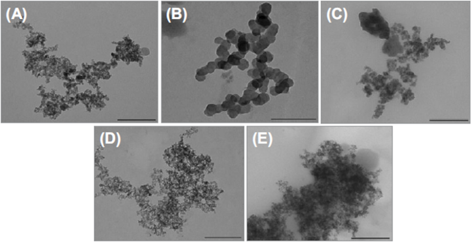
Mesoporous particles for the development of drug delivery system safe to human bodies

**************************************************************************

Atrazine—the turning point in Sexuality 

The herbicide atrazine is one of the most commonly applied pesticides in the world. As a result, atrazine is the most commonly detected pesticide contaminant of ground, surface, and drinking water. Atrazine is also a potent endocrine disruptor that is active at low, ecologically relevant concentrations. Previous studies showed that atrazine adversely affects amphibian larval development. The present study demonstrates the reproductive consequences of atrazine exposure in adult amphibians. Atrazine-exposed males were both demasculinized (chemically castrated) and completely feminized as adults[F1] . Ten percent of the exposed genetic males developed into functional females that copulated with unexposed males and produced viable eggs. Atrazine-exposed males suffered from depressed testosterone, decreased breeding gland size, demasculinized/feminized laryngeal development, suppressed mating behavior, reduced spermatogenesis, and decreased fertility. These data are consistent with effects of atrazine observed in other vertebrate classes. The present findings exemplify the role that atrazine and other endocrine-disrupting pesticides likely play in global amphibian declines.

Atrazine is one of the most widely used pesticides in the world. Approximately 80 million pounds are applied annually in the United States alone, and atrazine is the most common pesticide contaminant of ground and surface water (1). Atrazine can be transported more than 1,000 km from the point of application via rainfall and, as a result, contaminates otherwise pristine habitats, even in remote areas where it is not used (2, 3). In fact, more than a half million pounds of atrazine are precipitated in rainfall each year in the United States (2).[F2] 

In addition to its persistence, mobility, and widespread contamination of water, atrazine is also a concern because several studies have shown that atrazine is a potent endocrine disruptor active in the ppb (parts per billion) range in fish (4, 5), amphibians (612), reptiles, and human cell lines (5, 1315), and at higher doses (ppm) in reptiles (1618), birds (19), and laboratory rodents (2028). Atrazine seems to be most potent in amphibians, where it is active at levels as low as 0.1 ppb (610). Although a few studies suggest that atrazine has no effect on amphibians under certain laboratory conditions (29, 30), in other studies, atrazine reduces testicular volume; reduces germ cell and Sertoli cell numbers (11); induces hermaphroditism [F3] (6, 8, 10); reduces testosterone (10); and induces testicular oogenesis [F4] (79, 31). Furthermore, atrazine contamination is associated with demasculinization and feminization of amphibians in agricultural areas where atrazine is used (32) and directly correlated with atrazine contamination in the wild [F5] (7, 9, 33, 34).

Despite the wealth of data from larvae and newly metamorphosed amphibians, the ultimate impacts of atrazine’s developmental effects on reproductive function and fitness at sexual maturity, which relate more closely to population level effects and amphibian declines, have been unexplored. In the present study, we examined the long-term effects of atrazine exposure on reproductive development and function in an all-male population of African clawed frogs (Xenopus laevis), generated by crossing ZZ females (sex-reversed genetic males) to ZZ males (SI Materials and Methods). The advantage of using this population is that 100% of the animals tested were genetic males. As a result, all hermaphrodites and females observed are ensured to be genetic males that have been altered by endocrine disruption. We examined sex ratios, testosterone levels, sexual dimorphism, reproductive behaviors, and fertility in males exposed to 2.5 ppb atrazine throughout the larval period and for up to 3 years after metamorphosis-- Results

Feminization.

All of the control animals reared to sexual maturity (n = 40) were males, on the basis of external morphology, whereas only 90% of the atrazine-treated animals (36 of 40) appeared male at sexual maturity (on the basis of the presence of keratinized nuptial pads on the forearms and the absence of cloacal labia). The other 10% of atrazine-exposed animals (n = 4) lacked visible nuptial pads on the forearms and had protruding cloacal labia, typical of females (Fig. 1). Upon dissection of two of the apparent females and laparotomy in another two, we confirmed that animals with cloacal labia were indeed females from the present study, on the basis of the presence of ovaries (Fig. 1F). To date, two atrazine-induced females have been maintained, mated with control males (Fig. 1G), and produced viable eggs (Fig. 1H). The resulting larvae were all male when raised to metamorphosis and sampled (n = 100), confirming that atrazine-induced females were, in fact, chromosomal males. Furthermore, atrazine-induced females lacked the DM-W further confirming that these atrazine-induced females were indeed chromosomal males [F6] --These ZZ females expressed gonadal aromatase[F7] , as did true ZW females (n = 4, from our stock colony), but ZZ males (n = 8, control or treated)-- Atrazine-induced females expressed aromatase in their gonads. (Top) DMRT-1 and DM-W genes from a representative control and an atrazine-exposed adult male (M) and female (F). Morphologic sex was assigned on the basis of the presence of testes (males) or ovaries (females). (Middle and Bottom) Cyp-19 aromatase expression from gonads of the same animals genotyped at Top, along with the control gene, rpL8--Atrazine decreased androgen-dependent sperm production, mating behavior, and fertility. (A and C) Largest testicular cross-sections for representative control (A) and atrazine-exposed males (C) from 2007. (B and D) Magnification of individual tubules for control (B) and atrazine-exposed (D) males. Arrowheads in B and D show outline of tubules. Control tubules are typically filled with mature spermatozoa bundles, whereas the majority of tubules in atrazine-exposed males lack mature sperm bundles and are nearly empty, with only secondary spermatocytes (SS) along the periphery of the tubule. (E) Fertility for control (Con) and atrazine-exposed (Atr) males. Pooled data from both 2007 and 2008 study are shown. *P < 0.005 (ANOVA). (F) Fertility plotted against sperm content (percentage of tubules with mature sperm bundles) for control males (black symbols) and atrazine-exposed males (red symbols) for the 2007 (circles) and the 2008 (squares) studies. Dashed lines indicate the lower limit for controls for fertility and sperm content. Sample size differs from the number of trials because no data are available from females that did not lay eggs--Previous studies showed that atrazine demasculinizes (chemically castrates) and feminizes exposed amphibian larvae, resulting in hermaphrodites (8, 10) or males with testicular oocytes (7, 9) at metamorphosis. Since our initial publications (7, 9, 10), the effects of atrazine on amphibian development and the significance of these effects to amphibian declines have been a subject of debate (30, 35, 36). Although some investigators, including Carr et al. (6), reported statistically significant effects of atrazine on gonadal morphology in X. laevis (P < 0.0003 for multiple testes and P = 0.0042 for hermaphrodites), others, using different experimental conditions and different populations of the same species, suggested that atrazine had no effect (29). Essential to this debate, however, is (i) the terminology used to describe gonadal abnormalities; (ii) the expertise and ability of other researchers to recognize abnormalities; (iii) the possibility of natural variation in sex differentiation processes between species and even between populations (or strains) within a species (37); and (iv) the long-term consequences and significance of the observed abnormalities to amphibian reproductive fitness. Here we describe complete and functional female development in genetic (ZZ) males exposed to atrazine, not the production of hermaphrodites or males with testicular oocytes. Thus, there is no confusion in the present study regarding proper terminology or proper identification. Furthermore, because we used an all genetic (ZZ) male colony and genotyped the atrazine-induced ZZ females, there is no question that atrazine completely sex-reversed genetic (ZZ) males, resulting in reproductively functional females. --- The present study thoroughly examines the long-term effects of atrazine on reproductive function in amphibians. Although a single published study attempted to examine long-term reproductive effects of atrazine in amphibians (38), the authors did not report examinations of morphology. Furthermore, their examination of fertility and breeding of atrazine-exposed males was conducted after animals were injected with reproductive hormones (human chorionic gonadotropin, hCG), effectively providing “hormone replacement therapy and reversing the effects of atrazine. The present study represents a more thorough examination of the effects of atrazine on sex hormone production, testosterone-dependent development and morphology, male reproductive behavior, and fertility-- Transcription and subsequent translation of aromatase leads to estrogen production, which in turn directs differentiation of the ovary from the undifferentiated gonad. Just as exogenous estrogen results in the differentiation of ovaries in exposed genetic (ZZ) male X. laevis (45), induction of aromatase and subsequent estrogen production likely explain the complete feminization of genetic male-- Furthermore, why only some males (10% in the present population) are completely feminized, whereas their siblings are merely demasculinized, remains to be explored--Furthermore, sex-reversed males (ZZ females) are only capable of producing genetic male (ZZ) offspring, so the sex ratio in exposed populations would be skewed both by the production of atrazine-induced ZZ females as well as by the fact that ZZ females can only produce ZZ (genetically male) offspring-- In fact, mathematical models suggest that this very mechanism (the production of sex-reversed all male-producing animals) could drive populations to extinction

 Where is Atrazine used

Atrazine is used on crops such as sugarcane, corn, pineapples, sorghum, and macadamia nuts, and on evergreen tree farms and for evergreen forest regrowth. It has also been used to keep weeds from growing on both highway and railroad rights-of-way- Atrazine is used specifically to control broadleaf weeds (such as milkweed, thistle, or pigweed)

***************************************************************************** [F8]  

Treatment of Organophosphate Exposure

In my experience, organophosphate exposure causes more than just the neurological problems associated with anticholinesterase activity. However, the medical literature, most of the time, concentrates on that aspect of the poisoning. Particularly in chronic poisoning, the chemical dissociates within the body, resulting in metabolites of the organic component (which appear to have slightly different properties from the original at first exposure), phosphorus (which is highly toxic and corrosive in its own right) and whatever the associated radical/chemical add-on which the manufacturers add (in the Gulf War, this appears to have been fluorine which is also toxic in its own right).* With each of these component parts, there are separate sets of symptoms, separate storage sites, separate means of elimination. When the chemicals are stored in the body, they can be mobilized back into the bloodstream (in greater concentrations than at the time of absorption) as a result of exercise, heat, emotional excitement/distress, infection, massage, exposure to nutrients via dermal route near the storage sites, or due to displacement by other chemicals/drugs to which the person is exposed. Any detox must therefore be carried out with considerable care and caution, to avoid overwhelming the body’s coping mechanisms because of overload by chemicals re-emerging into the bloodstream.

There is precious little about chronic phosphate poisoning in modern literature; however, older books do deal with phosphorus poisoning because it was common in people manufacturing matches at that time. Body fluids containing phosphorous (particularly feces, urine, and possibly semen) will burn. Phosphorus bind strongly to mucous membranes (the cause of diarrhea and watery eyes in GWI cases). The toxic effects of chemicals can be somewhat ameliorated by ingestion of calcium acetate or calcium carbonates for phosphate binding and magnesiumand iodine for flourines – it binds to phosphorous and the organics, preventing some of the caustic effects, and helping prevent re-absorption back into the system through the intestinal wall. Both phosphorus and fluorine can be bound by calcium and magnesium. A combination of calcium citrate and magnesium oxide, in approximate physiological balance, plus potassium citrate or chloride, can be taken as needed throughout the day as symptoms are noted in the muscles, head, extremities.

*****************************************************************

Remedy

What you will need is

Sunflower Lecithin-calcium in some form ( acetate-carbonate-chloride) magnesium( citrate-malate-chloride any one of these) potassium (chloride or citrate either one) copper –selenium and zinc mixed using 1 gram increments with the minerals ( with the exception of selenium use 1 mg( milligram) and mix 2 oz of the liquid sunflower lecithin or add ( sunflower lecithin) powder ( egg yolk lecithin would be another alternative ) never use the soy lecithin- mix them all til they are completely saturated into the SFL—this will form a liposome---and then use 1 tsp several times a day---this will flush out the glyphosates and may cause a diarrhea effect –due to the flushing and restore the colon as well with probiotic or fermented foods

Because the organophosphates diminish absorption of nutrients while increasing the requirement for them, both this preparation, plus the calcium/magnesium and potassium mentioned above, can be dissolved in the mouth – absorbing through the mucous membranes of the mouth[F9] . Individual supplements can be taken as desired –, copper (which phosphorus binds to), zinc (balances copper), Vitamin C is also useful,– both from The corrosive effect of the phosphorus, and from the storage of fluoride which makes teeth and bones brittle. Dissolved Green Multi dissolves through the skin, too. --All fluorine exposure should be avoided – which may be difficult considering water supplies are being increasingly fluoridated. Fluoride absorbs through the skin as well as via mouth, making hot tubs and swimming pools places to add to the body's already excessive fluoride.

The old standby for restoring damaged myelin sheaths around nerves – lecithin – contains a whopping amount of phosphorus. However, some of it may have to be taken in order to restore neurological integrity or prevent further damage. Taking choline and inositol separately via supplement appears to dislodge stored chemicals from brain and other neurological tissue all at once, creating some rather bizarre and unsettling effectsDuring the process of detox and mending, the body's detox and eliminatory systems need to be supported.. It needs to be noted that clothing contaminated with organophosphates cannot be washed clean, and trying to wash it with other items of clothing results in the contamination of all of them. Storage in drawers and closets will also result in contamination not only of other clothing items in the same place, but also contaminate walls, floors of closets, and the wood in the chests of drawers. Contaminated walls can be washed down first with organic soap and then immediately with a slurry of lime. This does not eliminate the chemical from the walls entirely, but creates a film which can bind to at least some of the chemicals as they out-gas. It will have to be repeated. While performing such an operation, vigorous fresh-air ventilation is essential. Protective clothing and face covering (disposable) would be helpful. Even with these precautions, there is no guarantee that such cleaning won't expose the person doing the cleaning to enough chemicals to cause discomfort or a few days of symptoms.

Sources

Phosphorus: Henniger, Gordon R., Chapter 4, Drug and Chemical Injury – Environmental Pathology, pp. 146-245 in Kissane, John M., ed., Anderson’s Pathology – 9th ed. (St. Louis, C.V. Mosby Co. 1990)

Merck Manual – 13th ed., p. 1977

McGuigan, Hugh Alister, Applied Pharmacology, pp. 743-746, (St. Louis, C.V. Mosby Co. 1940)

Fluorine: Smith, Frank A., Fluroide Toxicity, pp. 277-283 in Corn, Morton, editor, Handbook of Hazardous Materials (San Diego, Academic Press 1993)

Hennigar, Gordon R., Chapter 4, Drug and Chemical Injury – Environmental Pathology, pp. 146-245 in Kissane, John M., ed., Anderson’s Pathology – 9th ed. (St. Louis, C.V. Mosby Co. 1990)

Merck Manual – 13th ed., p. 1970

McGuigan, Hugh Alister, Applied Pharamcology (St. Louis, C.V. Mosby 1940), p. 184

Organophosphates: Hennigar, Chapter (same as above), p. 226

Berkow, Robert, ed., The Merck Manual of Diagnosis and Therapy – 13th ed. (Rahway, NJ, Merck, Sharp & Dohme Research Laboratories 1977), p. 1482.

Dreisbach, Robt. H. and Wm. O. Robertson, Handbook of Poisoning: Prevention, Diagnosis and Treatment (Appleton & Lange, Norwalk, CT 1987) pp. 110-118.

*************************************************************************

Scientists Bioengineer First Artificial Animal Limb

Rat forelimb designed and grown in lab first step toward producing parts for transplant, researchers say


By Margaret Farley Steele

Thursday, June 4, 2015

THURSDAY, June 4, 2015 (HealthDay News) -- Researchers say they have taken the first steps in creating artificial replacement limbs that could be used for transplantation in the future.-Massachusetts General Hospital scientists have developed bioartificial rat forelimbs with functioning vascular and muscle tissue. (The term "bioartificial" means using biomaterials and cells to make replacement organs or limbs.) [F10] The scientists believe this process could one day work in primates -- including humans -- as well.-The researchers reported their findings online recently in the journal Biomaterials.-Although advances in prosthetic technology have helped the more than 1.5 million U.S. residents who are missing limbs, the artificial devices have functional and aesthetic drawbacks, the study authors noted. And transplants with donor hands, developed over the past two decades, expose recipients to risky immunosuppressive therapy for life, the researchers said.-"The composite nature of our limbs makes building a functional biological replacement particularly challenging," said study senior author Dr. Harald Ott. -"Limbs contain muscles, bone, cartilage, blood vessels, tendons, ligaments and nerves -- each of which has to be rebuilt and requires a specific supporting structure called the matrix. We have shown that we can maintain the matrix of all of these tissues in their natural relationships to each other, that we can culture the entire construct over prolonged periods of time, and that we can repopulate the vascular system and musculature," Ott said in a hospital news release.-Ott and his colleagues explained that a recipient can provide the specific cells needed to regenerate all of the tissues that make up a limb. "What has been missing is the matrix or scaffold on which cells could grow into the appropriate tissues," they said in the news release. -In Ott's previous research, living cells were stripped from a donor organ with a detergent solution. The remaining matrix was repopulated with specific cells appropriate to the particular organ.-For the new study, he and his colleagues used this "decellularization" technique to tackle the complexities of a bioartificial limb, while growing muscle and vascular cells in culture. -According to the news release, the resulting limb is complete with vascular cells along blood vessel walls and appropriate fibers of muscle cells.-When transplanted into animals, blood circulated through the vascular system, and electrical stimulation of muscles flexed the wrists and digits of the animals' paws, the researchers said. -Although animal research isn't necessarily applicable to humans, the researchers said they also successfully decellularized baboon forearms to show the feasibility of working on a human scale.-One of the upcoming challenges, Ott said, is regrowing nerves within a limb graft and reintegrating them into a recipient's nervous system. "Additional next steps will be replicating our success in muscle regeneration with human cells and expanding that to other tissue types, such as bone, cartilage and connective tissue," he said.-SOURCE: Massachusetts General Hospital, news release, June 2, 2015

****************************************************************************

Blood pressure medications can lead to increased risk of stroke

Date:May 29, 2015

Source:University of Alabama at Birmingham

Summary:The importance of preventing hypertension is reinforced by a study showing anti-hypertension medicines can increase stroke risk by 248 percent, according to new research. -- University of Alabama at Birmingham. "Blood pressure medications can lead to increased risk of stroke." ScienceDaily. ScienceDaily, 29 May 2015. <www.sciencedaily.com/releases/2015/05/150529193554.htm>.--Untreated high blood pressure, or hypertension, wreaks havoc on the body, leading to heart disease and stroke. New research from the University of Alabama at Birmingham published in the journal Stroke shows that, although HBP medications are beneficial, it is as risky to wait for the condition to develop and then treat it to a controlled level.--A cohort of 26,785 black and white participants ages 45-plus from the Reasons for Geographic and Racial Differences in Stroke (REGARDS) study were followed for 6.3 years by a research team led by George Howard, Dr.P.H., a professor in the Department of Biostatistics in the UAB School of Public Health. At baseline, 12,327 participants were successfully treated hypertensives, meaning their HBP treatment had their systolic blood pressure < 140 mm HG, the goal level set by the American Heart Association, and 4,090 unsuccessfully treated hypertensives.--At the conclusion of the follow-up period, more than 820 participants had experienced a stroke. -The harder hypertension is to control, the higher the risk for stroke, even if the treatment is successful. Howard says the risk of stroke went up 33 percent with each blood pressure medicine required to treat blood pressure to goal. Compared to people with systolic blood pressure below 120 mmHg without treatment, hypertensive individuals on three or more blood pressure medications had a stroke risk of 2.5 times higher.-"You're in as much trouble by the time you are on three medications that achieve excellent control as you are when you have hypertension and it is untreated, which is amazing," [F11] Howard said. "We want to raise the issue that, despite great advances in a pharmaceutical approach, relying solely on this approach is going to come at a dear price of people's lives."-The way to curb the problem, Howard says, is to prevent hypertension in the first place. There are a number of proven approaches to prevent or greatly delay the development of hypertension including: 1) taking part in moderate physical activity, 2) keeping weight in normal rages, 3) eating a diet rich in fruits, vegetables and Full fat dairy products and reduced in margarines-soybean oils-vegetable oils and canola and sugars-grains-breads-pastas-rice-soy and corn-eliminate the microwave.--"It's everything we know we should be doing," Howard said. "And over the past 14 years, stroke deaths are down 42 percent, likely because of this general shift of everybody in the population working toward having lower blood pressures."--Howard suggests that some future efforts to prevent the development of hypertension will have to be made through policy changes, such as increasing activity and limiting body weight require individual commitment.-"We need to keep the pressure to keep good things happening from a policy standpoint," Howard said. "Also, as individuals, we need to take the right actions for our health. Individuals and society need to work together to keep people from becoming hypertensive."-Study co-authors include Virginia J. Howard, Ph.D., professor in the Department of Epidemiology, Paul Muntner, Ph.D., professor and vice chair in the Department of Epidemiology, and Suzanne Oparil, M.D., professor in the Division of Cardiovascular Disease.--Story Source-The above story is based on materials provided by University of Alabama at Birmingham. The original article was written by Nicole Wyatt. Note: Materials may be edited for content and length.

***************************************************************************

Life-prolonging protein could inhibit aging diseases

Date:May 28, 2015

Source:Waseda University

Summary:Researchers have found a molecule that plays a key link between dietary restriction and longevity in mammals. This discovery may lead to the development of new therapies to inhibit age-related diseases. Waseda University. "Life-prolonging protein could inhibit aging diseases." www.sciencedaily.com/releases/2015/05/150528082026.htm>.--Researchers have found a molecule that plays a key link between dietary restriction and longevity in mammals. This discovery may lead to the development of new therapies to inhibit age-related diseases.--Studies have shown that moderate dietary restriction can increase the lifespan of many organisms, including mammals, while inhibiting age-related diseases such as cancer and Alzheimer's.-A team of researchers from Japan and Korea set out to understand how this happens. They discovered that a protein, called neuropeptide Y (NPY), which transmits nerve impulses in the brain and autonomic nervous system, is an essential link between dietary restriction and longevity in mice.-The researchers found that dietary restriction did not increase the lifespan of mice devoid of NPY to the same degree that it did in wild mice that had this protein. Dietary restriction also did not have the same protective effect against tumour formation and stress that it did in wild mice.-On the other hand, other adaptive processes that are activated during dietary restriction, such as changes in energy metabolism, were not affected by the absence of neuropeptide Y.--What's more, other researchers have found that an over-expression of the NPY gene increased the lifespan of rats that were not exposed to dietary restriction.--The Japan-Korea team's research, published in Scientific Reports, suggests that activating certain organs or tissues -- such as the liver, heart, other muscles and blood vessels -- with NPY could play a role in preventing age-related disorders, including cancer and Alzheimer's.--The researchers are currently engaged in joint exploratory research with pharmaceutical and food companies to develop substances that increase the expression of NPY as a deterrent to age-related diseases. Neuropeptide Y molecule. This neurotransmitter peptide plays an important role in food intake.-Story Source-The above story is based on materials provided by Waseda University. Journal Reference- Takuya Chiba, Yukari Tamashiro, Daeui Park, Tatsuya Kusudo, Ryoko Fujie, Toshimitsu Komatsu, Sang Eun Kim, Seongjoon Park, Hiroko Hayashi, Ryoichi Mori, Hitoshi Yamashita, Hae Young Chung, Isao Shimokawa. A key role for neuropeptide Y in lifespan extension and cancer suppression via dietary restriction. Scientific Reports, 2014; 4 DOI: 10.1038/srep04517


***************************************************************************

Mesoporous particles for the development of drug delivery system safe to human bodies

National Institute for Materials Science. "Mesoporous particles for the development of drug delivery system safe to human bodies." ScienceDaily. ScienceDaily, 20 May 2015. <www.sciencedaily.com/releases/2015/05/150520151334.htm>.

 Date:May 20, 2015

Source:National Institute for Materials Science

Summary:Scientists have succeeded in developing porous particles (mesoporous particles) consisting solely of phospholipids, a biological component, that are suitable for use as a drug delivery system--A research group led by MANA Scientist Kohsaku Kawakami, postdoctoral researcher Shaoling Zhang and MANA Principal Investigator Katsuhiko Ariga, at the International Center for Materials Nanoarchitectonics (MANA), NIMS (Sukekatsu Ushioda, President), succeeded in developing porous particles (mesoporous particles) consisting solely of phospholipids, a biological component, that are suitable for use as a drug delivery system[F12] . This study had been published in the Journal of Physical Chemistry C on 16 March, 2015.--Mesoporous materials are a type of material capable of serving as a drug delivery system[F13] . In conventional studies, hard materials such as silica and carbon materials have been used for such purposes, posing safety concerns to patients. The mesoporous material developed in this study consists exclusively of biologically-derived materials and is therefore expected to be very safe for humans.--Acquisition of official approval is one of the hurdles in the development of materials for use as a drug delivery system. To develop a certified pharmaceutical product, it is necessary to demonstrate the safety of the additive to be used before investigating the safety of the product itself. For this reason, pharmaceutical companies tend to avoid using new additives, which had been slowing the development of new drug carriers. However, the phospholipids examined in this study have already been used as emulsions and liposomes, and thus are not regarded as new additives. This fact is a great advantage of this material in view of commercialization.--This material comprises highly uniform mesoporous particles with diameters ranging between 5 and 20 μm[F14] , depending on their composition. It is a very lightweight material with a bulk density of about 0.02 g/cm3, from which an aerodynamic diameter of 1 to 3 μm is calculated. These are ideal features for this material to be used as a powder inhalation carrier.--Since this material consists of lipid bilayer membranes that are similar to biological membranes, it possesses the characteristics of both mesoporous particles and liposomes. For example, it can be used with both hydrophobic and hydrophilic drugs. Hydrophobic drugs can be embedded in a lipid bilayer membrane, and hydrophilic drugs can be inserted into hydrophilic regions between lipid bilayer membranes. Furthermore, as it is also feasible for the material to hold drugs in its mesopores, the material is capable of carrying drugs with various physical properties. Since phospholipids can be easily modified, it is conceivable that various kinds of surface modifications can be applied to the material.--This material is suited for industrial production as it can be easily prepared through freeze-drying. And it is expected to be useful as a drug carrier assuming any administration route and as a cosmetic ingredient. The unique shape of the particles also may add value to the commercial product.--Story Source-The above story is based on materials provided by National Institute for Materials Science. Journal Reference-Shaoling Zhang, Kohsaku Kawakami, Lok Kumar Shrestha, Gladstone Christopher Jayakumar, Jonathan P. Hill, Katsuhiko Ariga. Totally Phospholipidic Mesoporous Particles. The Journal of Physical Chemistry C, 2015; 119 (13): 7255 DOI: 10.1021/acs.jpcc.5b00159

 Top 


 [F1]Basically a homosexual effect in an amphibian

 [F2]And that is just in the USA not to mention Canada or Europe or Asia-Africa--etc

 [F3]Hermaphrodite—but male and female

 [F4]castration

 [F5]Sexual Alteration

 [F6]lesbianism

 [F7]Male Hormone

 [F8]This may also be true with other species when the endocrine system is off or altered it could carry over into the next generation

 [F9]Another method would be to mix in a yogurt or kefir utilizing the bacteria to break down these elements so that they are more useable to the system

 [F10]Potential Nano

 [F11]So taking the meds do not work

 [F12]This would be similar toa liposomal delivery method using a phosphorous based fat ( egg-sunflower lecithin-egg yolk lecithin) to transport neutrients through the body and cells

 [F13]Or vitamins or herbs or minerals

 [F14]Micron or micrometer which is safe to utilize

*******************************************************************************************************************************************8

Top A

Home


Script of the Show June 13 2015


Caffeine and gold to kill cancer cells

Low glycemic index diet reduces symptoms of autism in mice

Catalysts safely and effectively remove estrogenic compounds from wastewater

VERIFIED DECLARATION OF OBJECTION TO VIOLATIONS OF RELIGIOUS FREEDOM,                                                                                                      ESTABLISHMENT OF RELIGION, AND GENOCIDE

 GROUNDING THE HUMAN BODY TO NEUTRALIZE BIOELECTRICAL STRESS FROM STATIC ELECTRICITY AND EMFs

***************************************************************************

Caffeine and gold to kill cancer cells

March 3rd, 2014 | Author: Daniel

The side effects of consuming too much caffeine (e.g., agitation, rapid heartbeat and difficulty sleeping) are well known, but recent research has shown that stimulant also has its good side. You can kill cancer cells. Now, an international team of scientists has found that the combination of caffeine-based compound and a small amount of gold could someday be used as an anticancer agent.--Caffeine and certain compounds based on it have recently been in the spotlight as potential anticancer treatments. But it is clear that drinking liters of coffee, caffeinated sodas and energy drinks, is not the solution, and the caffeine in these drinks at the levels needed to kill cancer cells, they also begin to have negative effects on healthy. Meanwhile, gold can also kill cancer cells, but, like caffeine, can damage. Given all this, the team of Michel Picquet, Institute of Molecular Chemistry, University of Burgundy in Dijon, France, and Angela Casini, Institute of Pharmacological Research of the University of Groningen in the Netherlands, decided to put the two together in certain configurations, to see if the new compounds based on caffeine and gold can act selectively against cancer cells, preventing growth without damaging the other.-For this purpose a series of seven prepared new compounds in the laboratory and studied. The scientists found that, in certain concentrations, one of the series compounds selectively killed human ovarian cancer cells without harming healthy cells. Furthermore, the compound acted specifically against architecture of DNA that is associated with cancer.-In the research and development have also participated Loic Stefan, Marc Pirrotta, David Monchaud, Ewen Bodio, Philippe Richard and Pierre Le Gendre, Institute of Molecular Chemistry, University of Burgundy in Dijon, France, Elena Warmerdam, Marina H. de Jager and Geny MM Groothuis, Institute of Pharmacological Research of the University of Groningen in the Netherlands, and Benoit Bertrand of both institutes.

**********************************************************************

Low glycemic index diet reduces symptoms of autism in mice

Date-June 9, 2015

Source-Salk Institute

Summary-The number of people diagnosed with autism -- a spectrum of disorders characterized by social avoidance, repetitive behaviors and difficulty communicating -- has risen dramatically over the past two decades for reasons that are unclear. A diet recommended for diabetics ameliorated signs of autism in mice, researchers have found. Although preliminary and not yet tested in humans, the findings might offer clues to understanding one potential cause of autism. ---The brains of mice fed a high glycemic index diet have greater numbers of activated immune cells (shown in red and green) called microglia.---Bread, cereal[F1]  and other sugary processed foods cause rapid spikes and subsequent crashes in blood sugar. In contrast, diets made up of vegetables, fruits are healthier, in part because they take longer to digest and keep us more even-keeled.---New research in a mouse model of autism showed that such low glycemic index diets, similar to the plans that people with diabetes follow to keep their blood sugar in check, reduced symptoms of the disorder in mice. Although preliminary and not yet tested in humans, the findings, published June 9 in the journal Molecular Psychiatry, might offer clues to understanding one potential cause of autism.--The number of people diagnosed with autism -- a spectrum of disorders characterized by social avoidance, repetitive behaviors and difficulty communicating -- has risen dramatically over the past two decades for reasons that are unclear.--More people may be diagnosed due to a broader definition of autism and better efforts in diagnosis, but a true increase in the disorder cannot be ruled out, according to the U.S. Centers for Disease Control. Lifestyle change is one potential factor out of many possible causes of autism.--'One thing that's driving a lot of general physiological changes in people is changes in the diet,' says the study's corresponding author Pamela Maher, a senior staff scientist in the laboratory of professor David Schubert at the Salk Institute for Biological Studies.--In the new study, the Salk scientists used a mouse model of autism -- an inbred strain of mouse previously found to display autism-like symptoms -- to ask whether lowering the level of dicarbonyl methylglyoxal (a common byproduct of sugar metabolism) could alleviate symptoms of autism in the animals.--The scientists fed pregnant mice either the high or low glycemic index diet and kept their offspring on the same diet after birth and weaning, because their brains are still forming crucial connections.--The researchers then used a battery of behavioral and biochemical tests to study the mice after weaning. The two groups of animals consumed the same number of calories and were identical in weight. But mice that ate a high-glycemic index diet showed all of the expected behavioral symptoms of autism[F2] . Their social interactions were impaired, they repeated actions that served no apparent purpose, and they groomed extensively.

The mouse models of autism on a normal lab diet (with a medium glycemic index) are already known to generate fewer new neurons, and some of their existing cells and neuronal connections are abnormal compared with those of normal mice.--Intriguingly, in the new study, the brains of mice modeling autism that were fed the high-glycemic index diet had drastically less doublecortin, a protein indicator of newly developing neurons, compared to predisposed mice on the low-glycemic index diet. The deficiency was especially obvious in a part of the brain that controls memory.--In addition, the brains of the high-glycemic index diet mice appeared to have greater numbers of activated microglia, the resident immune cells of the brain. Their brains also expressed more genes associated with inflammation, compared to the mice fed the low-glycemic index diet.---Other studies of human mothers and their children with autism have implicated the activation of the immune system. For the most part, these studies have focused on infection, which causes a bout of inflammation -- as opposed to a high-glycemic index diet, which causes chronic, low-level inflammation, Maher says.--The new study found that the diet might directly influence the ecosystem of bacteria in the gut. More complex starches are broken down by bacteria that live in the lower part of the gut, the large intestine. The group saw some evidence of that in the blood, detecting metabolites that could only have come from the gut in larger amounts in the animals fed the high-glycemic index diet.--'We were really surprised when we found molecules in the blood that others had reported could only be generated by gut bacteria,' Maher says. 'There were big differences in some of these compounds between the two diets.'--The group plans to analyze the gut bacteria, and its potential link with features of autism, more directly. They also hope to better understand the role of inflammation in the ability to generate new neurons.--Lastly, they plan to vary the timing of exposure to the various diets in the mouse model of autism, by, for example, giving pregnant mice a high-glycemic index diet and then keeping their pups on a normal diet.--Story Source-The above story is based on materials provided by Salk Institute. -Journal Reference-A Currais, C Farrokhi, R Dargusch, M Goujon-Svrzic, P Maher. Dietary glycemic index modulates the behavioral and biochemical abnormalities associated with autism spectrum disorder. Molecular Psychiatry, 2015; DOI: 10.1038/mp.2015.64

***************************************************************************

Catalysts safely and effectively remove estrogenic compounds from wastewater

Date-June 12, 2015

Source-Carnegie Mellon University

Summary-Catalysts effectively and safely remove a potent and dangerous endocrine disruptor from wastewater. Scientists have demonstrated that the catalysts could be a viable option for large-scale water treatment.--Catalysts created by Carnegie Mellon University chemist Terrence J. Collins effectively and safely remove a potent and dangerous endocrine disruptor from wastewater.--In a paper published in Scientific Reports, Collins' research team and collaborators led by Brunel University London's Susan Jobling and Rak Kanda demonstrate that the catalysts could be a viable option for large-scale water treatment.--As pharmaceutical use has skyrocketed, especially in first-world countries, the amount of drugs released into the water system through wastewater has dramatically increased. Medications designed to disrupt the endocrine system, such as birth control pills and some breast and prostate cancer drugs, can be found in close to 25 percent of the world's streams, rivers and lakes. Studies have shown that these compounds have an adverse effect on the health of wildlife.--In many cases, researchers are finding that male fish in these polluted water sources undergo a process called feminization, which is an indicator that estrogenic contaminants are present in the water[F3] . Prolonged exposure to these female hormones can cause males to develop eggs in their testes and leads to the decline of fish populations.--"Unfortunately, some synthetic chemicals, including some everyday chemicals, are powerful endocrine disruptors and they often turn up as contaminants in water. These chemicals, called micropollutants,[F4]  can be bioactive at low environmentally relevant concentrations and are typically tough to break down," said Collins, the Teresa Heinz Professor of Green Chemistry at Carnegie Mellon. "We need to get these micropollutants out of our water systems. Fish are indicators of what can happen when hormone control systems get hijacked by synthetic chemicals. We humans are also animals with endocrine systems, after all."--When a person takes a drug, that drug travels through their body and what isn't absorbed or broken down is excreted as waste[F5] . Conventional wastewater treatment systems are unable to fully remove many of the harmful chemicals found in today's pharmaceuticals, pesticides and other products[F6] . Advanced processes installed at the end of wastewater treatment plants, especially those that use ozone or activated carbon, have been shown to be effective options for reducing micropollutants,[F7]  but the high financial and energy costs of incorporating these have limited their adoption.--Collins has developed a group of catalysts called TAML activators that offer an alternative treatment option. TAMLs are small molecules that mimic oxidizing enzymes.[F8]  When combined with hydrogen peroxide, TAML activators very effectively break down harmful chemicals in water. To test the effectiveness and safety of these catalysts, Collins teamed up with the Brunel research team, who are world-class experts in aquatic toxicity and wastewater treatment. In the current paper, the group demonstrates the efficacy and safety of TAML activators via a series of experiments. First, they showed that TAMLs were able to degrade, in pure water, 17alpha-ethinylestradiol (EE2), a synthetic estrogen found in oral contraceptives and a major cause of fish feminization. They then isolated the early intermediate compounds created as TAMLs degrade EE2, and found that several of these were estrogenic and harmful, too. But, using chemical analysis, the researchers showed that the TAML process was able to effectively degrade these intermediate compounds.--The research group also applied TAML activators to samples of water processed by municipal wastewater plants from the U.K. They found that the TAMLs were able to break down EE2 and other estrogenic compounds and micropollutants in the water.--The researchers were then able to demonstrate in the lab that water treated with TAMLs was not harmful to fish. They exposed male fathead minnows, a common freshwater fish found in many inland waterways, to water containing EE2. The exposure to EE2 caused a well-known effect -- the fish began to feminize. After they used TAMLs to remove EE2 from the water, the amount of vitellogenin, a female egg yolk protein, found in the minnows significantly decreased, signaling a dramatic reduction in feminization. Additionally, the fish did not have any detectable adverse effects from being exposed to the tiny traces of TAMLs in the water.--The researchers plan to test TAMLs against ozone and activated carbon treatment systems. They have shown that TAMLs will be at least as effective, and anticipate that the TAML process will come at a much lower cost. Collins estimates that a kilogram of catalyst could treat tens of thousands of tons of wastewater.--Story Source-The above story is based on materials provided by Carnegie Mellon University. Journal Reference-Matthew R. Mills, Karla Arias-Salazar, Alice Baynes, Longzhu Q. Shen, John Churchley, Nicola Beresford, Chakicherla Gayathri, Roberto R. Gil, Rakesh Kanda, Susan Jobling & Terrence J. Collins. Removal of ecotoxicity of 17α-ethinylestradiol using TAML/peroxide water treatment. Scientific Reports, 2015 DOI: 10.1038/srep10511


*****************************************************************************

VERIFIED DECLARATION OF OBJECTION TO VIOLATIONS OF RELIGIOUS FREEDOM, ESTABLISHMENT OF RELIGION, AND GENOCIDE

1. I, _______________________________, being over the age of 18, having firsthand knowledge of the facts contained herein, and being competent to testify to these matters, declare as follows: 2. Title 21 of the U.S. Code, Section 321 of The Food, Drug, and Cosmetic Act, purports to be "law" expressly applying to People whom it describes by the phrases "man or other animals" and "animals other than man". These phrases deem, declare and/or presume Americans to be animals in violation of fundamental principles of the Abrahamic Faiths (Judaism, Christianity and Islam) and violate the Organic Laws of the United States of America; the Declaration of Independence, the Establishment Clause and Free Exercise Clause of the First Amendment to the Constitution for the United States of America, as well as Article 18 of the Universal Declaration of Human Rights and statutes found at 18 U.S.C. §§ 241-242. 3. As one of the People with a sincere faith in the existence of a Creator having endowed mankind with certain God-given unalienable Rights, pursuant to the Organic Laws of the United States of America and the Declaration of Independence, I am adversely affected,

VERIFIED DECLARATION OF LIVING MAN / WOMAN ­ Pg 1 --injured and/or damaged by any law, Act of Congress, and/or conspiracy which would deem me and/or fellow members of mankind and the People of The United States of America to be "animals". "MAN OR OTHER ANIMALS" LAWS 4. 21 U.S.C. § 321(g) defines the term "drug" in relevant part as "(A) ...; and (B) articles intended for use in the diagnosis, cure, mitigation, treatment, or prevention of disease in man or other animals; and (C) articles (other than food) intended to affect the structure or any function of the body of man or other animals..." [Emphasis added]. 5. Numerous other instances of the phrases "man or other animals" and/or "animals other than man" exist in The Food, Drug and Cosmetic Act, as well as in the food and drug laws of the several states. 6. The phrases "man or other animals" and "animals other than man" mean that all men to which such food and drug laws are intended to apply are declared and/or presumed to be "animals". 7. The declaration and/or presumption that all men are "animals" violate fundamental principles of religious freedom and God-given unalienable Rights; evades the solemn obligations of government and implicates genocide. RELIGIOUS FREEDOM 8. Regardless of one's faith or religion, one of the most fundamental principles upon which The Declaration of Independence was written is the undeniable and irrefutable existence of all that is, indicating a Creator as written in Genesis 1:1 which states: "In the beginning, God created the heavens and the earth." By virtue of this creative Act, the Creator, however regarded or worshipped by man, owns perfect title to that Creation. 9. A second fundamental principle is found at Genesis 1:26-29 which declares that on the sixth day, God created man, and man alone, in God's image, and gave man dominion over all the animals. Thus man, having been created in the image of the Creator and having been given dominion OVER ALL THE ANIMALS, cannot therefore be deemed an –

VERIFIED DECLARATION OF LIVING MAN / WOMAN ­ Pg 2 -"animal" by any law of any man. Any "man" who subjects himself to such a law or accepts the status of "animal" violates this most fundamental principle. 10. The U.S. Code defines the Organic Laws of the United States to include The Declaration of Independence, which states in part: "We HOLD these Truths to be self-evident, that all Men are CREATED equal, that they are endowed by their Creator with certain unalienable Rights..." 11. The term "hold" is a legal term with which the judiciary is well acquainted. It means to "preserve as a matter of law". It is held ­ preserved, as a matter of law, by the very Organic Laws of the United States of America, that "all men are created" and are "endowed by their Creator" ­ with certain God-given unalienable Rights. 12. As per Acts 17:26, all men are created from the same blood. No man can be treated as an animal. As per Acts 17:27, the Creator appoints to each man the task to "seek the [Creator] in the hope that they might find Him, though He is not far from each one of us..." 13. Any de jure or de facto government, governmental agency and/or corporation that acts to degrade any man created in the Creator's image to the status of an "animal" would trespass upon that man's religious freedom, interfere with that man's search for his Creator by implicitly denying the Creator's existence, and/or seek to subject that man to an establishment of a religion such as secular humanism, naturalism, or Darwinism. 14. The unanimous Declaration of the original thirteen united States of America, as well as Article 1, Section 11 of the Constitution of The State of Washington, and the First Amendment to The Constitution of the United States of America; each include fundamental principles, guarantees and/or prohibitions that secure each man's right of religious freedom and right to be free from the establishment of religion. These same instruments impose solemn obligations upon government to secure such rights and entitle every man to seek redress for violations by government of such obligations. 15. Any state or federal law that deems, declares, or presumes man to be an "animal" is anathema to my faith. Such laws constitute the establishment and imposition of a VERIFIED DECLARATION OF LIVING MAN / WOMAN ­ Pg 3 -religion of secular humanism, naturalism, or Darwinism upon the People of The United States of America. Any act by any state or federal government or governmental agency that directly or indirectly degrades mankind to the status of "animals" likewise constitutes the establishment of a religion of secular humanism, naturalism, or Darwinism upon the People of The United States of America. Such "man or other animals" law(s) are facially unconstitutional and unconstitutional as applied in the matter before this tribunal. 16. I hereby object to any law that declares or presumes me or my fellow man to be an "animal". As a matter of love for my neighbors, I object on behalf of all mankind and the People of The United States of America, to any such law that degrades the People to the status of "animals". 17. Any decision knowingly based on laws that presume and/or declare all men to be "animals" would constitute a conspiracy to violate and/or deny the God-given, unalienable Rights of the People of The United States of America to religious freedom and/or freedom from a government-imposed establishment of religion. Such a decision would further damage said People by depriving them of their standing as "sovereigns without subjects" and "joint-tenants in the sovereignty" as declared by The Supreme Court of the United States in Chisholm v State of Georgia, 2 Dall. 419 (A.D. 1793). 18. Parties to this matter may not have previously understood that The Food, Drug, and Cosmetic Act deprives the People of their God-given, unalienable Right to religious freedom and/or freedom from establishment of religion. Parties to this matter might previously have argued that they did not knowingly intend to deprive People of such rights. However, thanks to this Notice, all parties now know or have reason to know that such further action based upon laws repugnant to the Constitution would cause injury and/or damages to me and the People of The United States of America. IMPLICATIONS OF CONSPIRACY TO COMMIT GENOCIDE 19. The Wikipedia.com article entitled "Genocide" reports in part, "In 1996, Gregory Stanton, the president of Genocide Watch, presented a briefing paper called `The Eight

VERIFIED DECLARATION OF LIVING MAN / WOMAN ­ Pg 4 Stages of Genocide' at the United States Department of State. In it, Stanton suggests that genocide develops in eight (8) stages that are "predictable but not inexorable." Those eight (8) stages include: 1) Classification; 2) Symbolization; 3) Dehumanization; 4) Organization; 5) Polarization; 6) Preparation; 7) Extermination; and 8) Denial. 20. Stage #3 ("Dehumanization") is described in part as "One group denies the humanity of the other group. Members of it are equated with animals, vermin, insects or diseases." [Emphasis added]. 21. History tells us that African-Americans were enslaved based on the most evil of premises that they were not "men" but chattel (property or "animals"). History tells us they fought the civil rights movement in large measure to be recognized as "men". The Civil War and the deaths of 750,000 Americans followed, in part, from the horrible premise that some men were "animals". 22. History also tells us that during World War II, Jews suffered extermination at the hands of the Nazi's based on the most evil of premises that the Jews were "untermenschen" (less than men or "animals"). Nazis could slaughter even Jewish women and children because to them they were no more than "animals". Millions of men and women died, the world was plunged into its most tragic war, and the nation of Germany was nearly destroyed based in part on the horrible premise that some men were "animals" 23. More recent history tells us that in April of 1994, in the African country of Rwanda, nearly a million men, women and children of the Tutsis and Hutus were hunted with machetes like animals, without any regard to their humanity. 24. Those state or federal food and drug laws which declare or presume man to be an "animal" conform to Stage Three of "The Eight Stages of Genocide", namely "Dehumanization" and characterization as "animals". 25. The United Nations' Convention on the Prevention and Punishment of the Crime of Genocide, approved December 9th, A.D. 1948, declares in part, "genocide is a crime under international law" -- but that crime need not include the actual deaths of any victims. For example, Article 2 of said Convention declares in part, "... genocide means

VERIFIED DECLARATION OF LIVING MAN / WOMAN ­ Pg 5-any of the following acts committed with intent to destroy, in whole or in part, a national, ethnical, racial or religious group, as such: ... Causing serious bodily or mental harm to members of the group; (c) Deliberately inflicting on the group conditions of life calculated to bring about its physical destruction in whole or in part; ..." [Emphasis added]. 26. The state and federal "man or other animals" laws inflict "mental harm" and/or impose "conditions of life calculated to bring about the physical destruction in whole or in part" upon those sovereign People of The United States of America who are made in the Creator's image (as per Genesis 1:26-29) and "endowed by their Creator with certain unalienable Rights" (as per The unanimous Declaration of the thirteen united States of America of July 4th, A.D. 1776). This injury is achieved by refusing to recognize said People's direct relationship to the Creator by implicitly denying the existence of the Creator and by degrading the People from the status of men, whose rights flow from the Creator, to the status animals whose "rights" -- if any -- flow from the government. 27. Article 3 of said Convention declares, "The following acts shall be punishable: (a) Genocide; (b) Conspiracy to commit genocide; (c) Direct and public incitement to commit genocide; (d) Attempt to commit genocide; (e) Complicity in genocide." [Emphasis added]. 28. Article 4 of said Convention declares, "Persons committing genocide or any of the other acts enumerated in article III shall be punished, whether they are constitutionally responsible rulers, public officials or private individuals." 29. Until now, parties in this matter may have had no understanding of the genocidal implications of their contemplated actions and thus no intent to engage in genocide. However, thanks to this Notice -- which I provide as an act of love for my neighbors -- the parties now know or have reason to know the genocidal implications of contemplated action and/or conspiracy based on "man or other animals" laws. 30. Should said parties proceed in their contemplated actions and/or conspiracy, they do so knowing or having reason to know that that such conspiracy may be found to constitute

VERIFIED DECLARATION OF LIVING MAN / WOMAN ­ Pg 6 an act of genocide as defined by the United Nations' Convention on the Prevention and Punishment of the Crime of Genocide. THE POWER OF A JURY 31. John Jay was the first Chief Justice of the U.S. Supreme Court, and the first President after the American Revolution in 1789. He said: "The Jury has a right to judge both the law as well as the fact in controversy." 32. Samuel Chase, who was a signatory to the Declaration of Independence and Associate Justice of the U.S. Supreme Court in 1796 said: "The jury has the right to determine both the law and the facts." 33. Oliver Wendell Holmes, another U.S. Supreme Court Justice in 1902 said: "The jury has the power to bring a verdict in the teeth of both law and fact." See Homing v. District of Columbia, 138 (1920). 34. Harlan F. Stone, 12th Chief Justice of the U.S. Supreme Court in 1941 said: "The law itself is on trial quite as much as the cause which is to be decided." 35. Justice Stone also said: "If a juror feels that the statute involved in any criminal offence is unfair, or that it infringes upon the defendant's natural god-given unalienable or constitutional rights, then it is his duty to affirm that the offending statute is really no law at all and that the violation of it is no crime at all, for no one is bound to obey an unjust law." 36. The Supreme Court said in Marbury v. Madison (5US137, 176; 1803): "All laws which are repugnant to the Constitution are null and void." 37. In U.S. v Moylan (1969), the 4th Circuit Court of Appeals said "If the jury feels the law is unjust, we recognize the undisputed power of the jury to acquit, even if its verdict is contrary to the law as given by a judge, and contrary to the evidence." 38. Lysander Spooner said in The Right of Juries: "For more than six hundred years ­ that is, since Magna Carta, in 1215 ­ there has been no clearer principle of English or American constitutional law, than that, in criminal cases, it is not only the right and duty of juries to judge what are the facts, what is the law, and what was the moral intent of the

VERIFIED DECLARATION OF LIVING MAN / WOMAN ­ Pg 7 accused; but that it is also their right, and their primary and paramount duty, to judge the justice of the law, and to hold all laws invalid, that are, in their opinion, unjust or oppressive, and all persons guiltless in violating, or resisting the execution of, such law." 39. In the recent U.S. Supreme Court case Bond vs. United States, No. 09-1227 (2011), the Court said: "'An offence created by [an unconstitutional law],' the Court has held, `is not a crime.' Ex parte Siebold, 100 U.S. 371, 376 (1880). `A conviction under [such a law] is not merely erroneous, but is illegal and void, and cannot be a legal cause of imprisonment.' Id., at 376­377. If a law is invalid as applied to the criminal defendant's conduct, the defendant is entitled to go free ... In short, a law `beyond the power of Congress,' for any reason, is `no law at all.' Nigro v. United States, 276 U. S. 332, 341 (1928)." 40. Thomas Jefferson is attributed to saying "If people let the government decide what foods they eat and what medicines they take, their bodies will soon be in as sorry a state as are the souls of those who live under tyranny." 41. The U.S. Food and Drug Administration ("FDA"), together with the U.S. Department of Justice ("DOJ"), seeks to charge, indict, or convict Mr. Daniel Smith of Project GreenLife, and others, for alleged violations of The Food Drug and Cosmetic Act. Mr. Smith could face many years in prison if convicted. However, The Food, Drug, and Cosmetic Act exceeds Congress' enumerated powers because it violates the Establishment Clause and Free Exercise Clause of the First Amendment to Constitution for the United States of America, which states in pertinent part: "Congress shall make no law respecting an establishment of religion, or prohibiting the free exercise thereof;" 42. The Declaration of Independence states: "... that to secure these Rights, Governments are instituted among Men, deriving their just Powers from the Consent of the Governed, that whenever any Form of Government becomes destructive of these Ends, it is the Right of the People to alter or abolish it..." 43. It is the right of the jury to HOLD (preserve as a matter of law), that The Food, Drug, and Cosmetic Act by its own definitions, which deem, declare and/or presume Americans

VERIFIED DECLARATION OF LIVING MAN / WOMAN ­ Pg 8 and all mankind to be merely "animals", violates God-given unalienable rights guaranteed by the Constitution for the United States of America, as well as Article 18 of the United Nations' Universal Declaration of Human Rights, which states: "Everyone has the right to freedom of thought, conscience and religion; ... and freedom ... in public or private, to manifest his religion or belief in teaching, practice, worship and observance. 44. It is the right of the jury to HOLD (preserve as a matter of law), that The Food, Drug, and Cosmetic Act by its own definitions, which deem, declare and/or presume Americans and all mankind to be merely "animals", conforms to Stage (3) Three of "The Eight Stages of Genocide" wherein "man" is characterized as an "animal". 45. In 1980, an American screenplay received eight (8) Academy Award nominations including "Best Picture". It depicted a man being chased on the streets of London, unmasked, and cornered by an angry mob, at which point he cries out before collapsing... "I AM NOT AN ANIMAL ... I AM A HUMAN BEING ... I ... AM ... A ... MAN". This screenplay was an adaptation from the books by Sir Frederick Treves and Ashley Montagu... The Elephant Man: A Study in Human Dignity. 46. The Food, Drug, and Cosmetic Act evades all manner of human dignity by deeming, declaring and/or presuming mankind to be merely "animals". We the People must stand up ­ for human dignity and for God-given unalienable Rights guaranteed by the Constitution for the United States of America, and to remind the Government that "Governments are instituted among Men, deriving their just Powers from the Consent of the Governed," and "that whenever any Form of Government becomes destructive of these Ends, it is the Right of the People to alter or abolish it." 47. Consistent with my faith, State Constitutions, and with the Organic Laws of the United States, including the Declaration of Independence and the Constitution for the United States of America and Article 18 of the Universal Declaration of Human Rights, I declare and give notice of the assured value and preservation of my God-given, unalienable Rights, my right to religious freedom, my right to be free from the establishment of religion, and that I and my fellow man, are NOT, nor ever shall be "animals".

VERIFIED DECLARATION OF LIVING MAN / WOMAN ­ Pg 9


48. If we are prepared to accept the label of "animal", we will be treated like animals. The government derives its power from the consent of the governed and I, for one, do not consent. I AM NOT AN ANIMAL! 49. Based upon the foregoing, the jury should exercise its right NOT to charge, indict or convict Daniel Smith, and should effectively return an acquittal - or a NO TRUE BILL OF INDICTMENT. The jury should use its unique and unequivocal Power to ferret out real crimes committed by real criminals beginning with government agencies like the FDA who seem to be perpetually engaged in the intentional abrogation of these, our Godgiven, natural, and unalienable rights.


VERIFICATION


I, ________________________________, hereby verify, under penalty of perjury, under the laws of the United States of America that the above statement of facts and laws is true and correct, according to the best of my current information, knowledge, and belief, so help me God, pursuant to 28 U.S.C. § 1746(1). Dated this _________ Day of _____________, 2012 A.D.


Signed Name: _______________________________________ Printed Name: _______________________________________ Address: _________________________________________ City, State, Zip: ______________________________________


VERIFIED DECLARATION OF LIVING MAN / WOMAN ­ Pg 10


***************************************************************

 GROUNDING THE HUMAN BODY TO NEUTRALIZE BIOELECTRICAL STRESS FROM STATIC ELECTRICITY AND EMFs.

A. Clinton Ober Ventura, CA INTRODUCTION


From the beginning of time, except for the past few generations, humans lived their entire lives primarily in direct physical contact with the earth; therefore, it is assumed that humans throughout evolution were naturally grounded. In modern times, humans have insulated themselves from contact with the earth by wearing synthetic soled shoes and living in homes that elevate the body above the earth. Consequently, humans are no longer naturally grounded and now the body becomes charged with static electricity and radiated electric fields can now create unnatural weak electric currents with-in the body[F9] . [1] This work provides evidence that loss of natural ground allows extraneous electricity to interfere with and stress the normal bioelectrical activities of the body, which thereby interferes with natural health and sleep. Today everyone is physically stressed, their muscles are tense, back and joint pain are the norm and most do not sleep well. These conditions all relate to excess stimulation of the nervous system and/or interference of the bioelectrical communications between cells. For instance, muscles only respond to bio-electrical communications from nerves. When these communications are interfered with muscles become tense and remain tight. This leads to fatigue, skeletal problems and pain. To what extent do EMFs create abnormal electrical activity in/or on the body? In 1995 the National Institute of Environmental Health Sciences [NIEHS] and the US Department of Energy [DOE] stated that common exposure to electric and magnetic fields [EMFs] from household electrical wires now produce unnatural weak electric currents between human cells. In other words 24 hours a day if you live and sleep in a modern home. [1] These unnatural currents in the body are the direct result of the body being insulated from ground contact[F10] . The question is do these currents along with the static electricity created on the body from carpets etc. interfere with normal bioelectrical functions? An indication is; according to the American Institute of Stress, over 75% of all visits to primary care physicians are now for stress related health conditions. The description of stress is; a state of continuous anxiety and nervousness in which muscles become and remain tensed. Stress is now confirmed to be a primary contributor to cardiovascular disease, cancer, gastrointestinal, skin, neurological and emotional disorders, and a host of disorders linked to immune system disturbances ranging from the common cold and herpes, to arthritis[F11] . [2] In the late 1960s, when humans were first widely diagnosed as being stressed, synthetic soled shoes, carpets and the like had just become popular and the use electricity and household electrical devices tripled from the previous generation. Do these unnatural weak electric currents in the body also interfere with sleep? According to the National Sleep Foundation's Sleep-2000 report [3], nearly two thirds of American adults [62%] now suffer from sleep problems. Americans have the most comfortable beds and the most protected sleep environments in the world. Yet, in traditional societies where most humans sleep on animal skins, grass mats or directly on the ground, sleep problems do not exist. [4] As for Americans, most now sleep within 12 inches of electrical wires hidden in the wall at the head of their bed and with electric cords around or near the bed. All of which emanate e-fields throughout the night and create weak electric currents in the body [1]. The fact that the majority of people, with the best health care in human history, now increasingly suffer from poor sleep and stress related health problems suggests that something, largely unknown to the health community and public, is wrong.[F12]  The dramatic change from the body being naturally grounded to now conducting unnatural weak electric currents between cells is the most likely candidate. Circumstantial evidence is provided by the fact that humans in traditional societies that maintain contact with the earth do not experience the common sleep and stress related health problems of the modern world [F13] [4]. Nor do the animals that live in direct contact with the earth. Evidence that is more conclusive was reported by the NIEHS and the DOE [1] that in some laboratory studies the biological effects of EMFs are: -Changes in functions of cells and tissue -Accelerated tumor growth -Decrease in the hormone melatonin -Changes in biorhythms -Alterations of immune system -Changes in human brain activity and heart rate The question is; by restoring natural ground to the body and thereby neutralizing these weak electric currents in the body and static electricity on the body, do muscle relax and normal sleep return? In search of the answer the following test was performed. METHOD AND MATERIALS -To effectively restore ground contact for an extended period, test subjects slept on dissipative carbon fiber mattress pads placed under their fitted sheets, connected via a ground wire [protected with an inline 1/100 amp fast blow fuse], to a ground rod driven into the earth near their bedroom window. The grounded mattress pads were designed to replicate the ground plane of the earth in the bed. Sleep disturbances along with chronic muscle and joint pain, which the subjects had been experiencing for at least six months, were recorded to establish a base line. The test was for a period of 30 days. SELECTION OF PARTICIPANTS An advertisement, distributed to ten beauty salons in Ventura County, CA solicited individuals experiencing sleep problems accompanied by tense muscles and/or chronic joint pain to participate in the study. Of the respondents, sixty individuals participated. Age of subjects was between 23 and 74 years Male subjects = 22 Female subjects = 38 Declared sleep problems = 100% Declared chronic muscle or joint pain = 100% The subjects were randomly divided into two groups. The first group of thirty, slept on carbon fiber mattress pads connected to a dedicated earth ground, just outside their bedroom window. The second control group of thirty, slept on carbon fiber mattress pads but were not connected to an earth ground. E-field created charge on their bodies were recorded with an AC voltmeter connected to the earth ground and body contact made with a hand held probe or an EKG electrode patch. E-field created charge measured on subject's bodies while lying in their beds were as follows: Test subjects Control subjects Less than 1 volt 2 2 1 volts or more 28 28 2 volts or more * 16 15 3 volts or more 8 6 4 volts or more 4 3 5 volts or more 3 2 *All subjects averaged 2+ volts on their bodies while lying in their beds. E-field created charge measured on test subject's bodies after grounding: averaged 10 millivolts or less.-

RESULTS

Categories: Time to fall asleep Quality of sleep Wake feeling rested Muscles stiffness & pain Chronic back and/or joint pain General well-being Test Subjects* Same Improved 4=15% 23=85% 2=7% 25=93% 0=0% 27=100% 5=18% 22=82% 7=26% 20=74% 6=22% 21=78% Control Subjects** Same Improved 20=87% 3=13% 20=87% 3=13% 20=87% 3=13% 23=100% 0=0 % 23=100% 0=0% 20=87% 3=13%


*Reports not received from three participants. **Reports not received from seven participants. DISCUSSION The purpose of this work was to provide evidence that when the human body is grounded it is naturally protected from static electricity and radiated electric fields. The meter reading of the grounded subject proved this true. The benefits of grounding the body were expected to relax muscles and improve sleep. This also proved to be true. Worthy of mention is that several subjects in the study stated they also experienced significant relief from asthmatic and respiratory conditions, rheumatoid arthritis, PMS, sleep apnea and hypertension, while sleeping grounded. These unexpected results indicate that loss of ground contact plays a much larger role in overall health then was anticipated at the start of this study. ADDITIONAL SUPPORT TO THESE FINDINGS In the May 1999 NIEH-EMF RAPID report, mention is made that reported biological effects to humans exposed to EMFs, such as changes in melatonin levels cannot be confirmed in animal studies. Therefore, the actual effects to humans are inconclusive. [5] In the animal studies, sheep exposed to EMFs from a power line were reported to have experienced no change in melatonin levels. The sheep, which walked and slept directly on the ground were naturally grounded throughout the experiment. The fact that melatonin levels remain normal in sheep when grounded, supports these findings that when humans are grounded their sleep improves.[F14] 

 Personal note from Roger Coghill, MA Biol. MI Biol. MA Environ Mgt. a leading research scientist and author specialized in the field of Bioelectromagnetics, the science investigating the interaction of electricity with organic life.--Yes, I would be prepared to believe that grounding the body will help dispel any extraneous electric fields which could otherwise interfere with the body's own endogenous fields. We have found that these latter are vital for wellbeing, with adverse effects if disturbed. It could also be a way forward for protection against high frequency radiation. Best, Roger Coghill 12/05/99- Comments  from Russell Whitten, D.C. Ojai, CA 8/25/00


Prompted by the results of a patient, who participated in Mr. Ober's study, I grounded the beds of 35 additional patients over a two-month period. The bed e-field measurements in this group ranged from .3 to 47 volts before grounding. A variety of health benefits occurred in this time. Many of the improvements, such as increased energy and athletic performance, can be attributed to the improved sleep that almost everyone reported. However, in many cases metabolic and hormonal conditions responded as well. Chronic back pain went away in several cases, stiff arthritic joints became more flexible, asthma attacks subsided and PMS symptoms lessened greatly. These indications confirm that e -fields do affect the body. CONCLUSIONS The important finding of this study is that the human body when grounded is naturally protected from static electricity and the weak electric currents created in the body by radiated electric fields. The benefits of grounding the body are; sleep significantly improves, muscles relax, chronic back and joint pain subsides and general health improves. PRINCIPAL REFERENCES 1. National Institute of Environmental Health Sciences and the U.S. Department of Energy, Questions and Answers about EMF, electric and magnetic fields associated with use of electric power [1995] 2. American Institute of Stress, www.stress.org/problems 3. National Sleep Foundation, www.sleepfoundation.org/pressarchives 4. Slumbers Unexplored Landscape [1999] Carol M. Worthman, Anthropologist, Emory University Atlanta, GA 5. National Institute of Environmental Health Sciences EMF Rapid Report [May, 1999] For additional information on this study or for parties interested in performing additional studies related to personal grounding and health, please contact: Clint Ober @ 805- 844-0888 or e-mail clintober @ prodigy.net


Top A


 [F1]These also have high levels of atrazine and glyphosates which have endocrine disrupting agents as well as digestion impairments that block crucial minerals and vitamins required for an effective brain health and function

 [F2]What you have to look at with this diet is what type of high glycemic diet

 [F3]Atrazine –Glyphosates and Pesticides are also hug contributors

 [F4]Or Xeno Sstrogens

 [F5]Pharma Copia-or Sorcers Death Construction

 [F6]The need for Distilled or RO water that does take these out

 [F7]Ozone and  Charcoal –Carbon---one oxidizes the other removes

 [F8]Tetra-amido macrocyclic ligands (TAMLs) constitute a class of synthetic molecules that serve as catalysts for oxidation reactions. They were first designed by Terrence J. Collins and associates at Carnegie Mellon University, and are touted as examples of environmentally friendly catalysts. Several variations exist, including an iron-TAML complex that can catalyze the breakdown of the pesticide fenitrothion. Other promising uses for iron-TAML catalysts include the treatment of toxic effluent streams from paper mills and eliminating corrosive dibenzothiophenes from diesel fuels. The potential use of TAML as a bactericide against anthrax spores, which are known for their resilience, is being investigated as well.

 [F9]There is the fact as well that with nano particles in the air being charged with RF’s and other Freq—and signals this to adds to the cellular and tissue loads

 [F10]This should really be saying with out an outlet to release the charge---if you have this you can dispel the charge as well and it is easy enough to do

 [F11]Here is where the article can be misleading ---Stress can be caused by another of things from being pulsed with frequencies to as well pathogens to pollutants to just having to deal with a matrix that is been built of deceptions—so can not being grounded cause stresss---would be more it can increase or exacerbate the condition more then cause

 [F12]Again it may have a amplifying effect of frequencies being pumped through the air at night between 3-3:30 coming from towers and  smart meters---so here again is where the need to use common sense and not allow the writer to dictate something on a premise

 [F13]That would be due to not be in a frequency smog filled environment ---but that is changing as well—people in the rural areas ae as well having difficulty sleeping and again it would be from another freq or charging effect ongoing

 [F14]Again this is speculation and not realistic—sheep are grounded all day people are not


**********************************************************************************************************************************************

TOP B

Home


Script of the Show June  20 2015

DMSO Therapy

**************************************************************************************************************************************************

DMSO Therapy

What It Does

DMSO tends to build up white blood cells and increase immune production of MIF (migration inhibitory factors) of macrophages. Thus, the immune system is made more effective by allowing macrophages to move more quickly. Thus DMSO modulates lymphocytes, and it therefore reactivates the production of MIF. It also diminishes allergic reactions by unfolding the cell membrane and making more cell receptor sites available to attachment by specific antigens. ----The modulating effect of DMSO on lymphocytes also tends to increase the production of lymphokines (chemical immune cell mediators) such as interferon. It potentiates cell mediated immunity and can be effective in multiple sclerosis, systemic lupus, erythematosus, rheumatoid arthritis, thyroiditis, ulcerative colitis, cancer, etc.

What Are Its Major Therapeutic Properties?

* It blocks pain by interrupting conduction in the small c-fibers, the non-myelinating nerve fibers.
* It is anti-inflammatory.
* It is anti bacterial, fungal and viral.
* It transports all molecules (drugs, etc.) across cell membranes.
* It reduces the incidence of platelet thrombi (clots in vessels).
* It effects cardiac contractility by inhibiting calcium to reduce the workload of the heart.
* It is a vasodilator, probably related to histamine release in the cells and to prostaglandin inhibition.
* It softens collagen.
* It is a scavenger of the hydroxyl free radical.
* It stimulates the immune system.
* It is a potent diuretic.
* It increases interferon formation.
* It stimulates wound healing.

Summary

DMSO has certain unique physiological characteristics which stem from its molecular makeup:

* It is a simple small molecule with unusual properties.
* An exothermic reaction occurs when DMSO is diluted with water (heat is generated).
* Hydroxyl radicals (OH), which are free radicals (oxidants), are ubiquitous and highly injurious to cells—and thus health. DMSO neutralizes (quenches) these free radicals. It is a free radical scavenger!
* DMSO substitutes for water in the living cell—it can destroy intracellular free radicals. No other antioxidant can do that.
* DMSO increases the permeability of cell membranes yielding a flushing effect of toxins from intracellular location to extracellular.
* It is an antidote to allergic reactions.
* It can penetrate any cell wall; thus it can get where most chemicals can’t.
* It has a very low index of any toxicity.
* Allergic reactions to DMSO can occur but they are uncommon.
* DMSO has a myriad of applications in medicine. Some are so dramatically effective that the concept of such therapy just boggles the mind!

References

Szmant, H. Harry. Physical properties of dimethyl sulfoxide and its function in biological systems, Biological Actions of Dimethyl Sulfoxide ed. by Stanley W. Jacob and Robert Herschler. (New York: New York Academy of Sciences, 1975), pp. 20-23.

Barfeld, H., and T. Atoynatan. N-acetylcysteine inactivates migration inhibitory factor and delayed hypersensitivity reactions. Nature new Bio., 231:157-159, 1971.

Barfeld, H., and T. Atoynatan, Cytophilic nature of migration inhibitory factor associated with delayed hypersensitivity, Proc. Soc. Exp. Biol. Med., 139:497-501, 1969.

Tschope, M., cited in Raettig, H. “The potential of DMSO in experimental immunology,” Dimethylsulfoxyl, Internationales Symposium in Wien. G. Laudahn and K. Getrich, eds.; 54. Saladruck, Berlin, Germany, 1966.

Engel, M.F. Ann. N.Y. Acad. Sci., 141:638, 1967.

 DMSO

Medical use of dimethyl sulfoxide (DMSO).

Swanson BN.

Abstract

DMSO is a clear odorless liquid, inexpensively produced as a by-product of the paper industry. It is widely available in the USA as a solvent but its medical use is currently restricted by the FDA to the palliative treatment of interstitial cystitis and to certain experimental applications. Cutaneous manifestations of scleroderma appear to resolve (albeit equivocally) following topical applications of high concentrations of DMSO. A limited number of small clinical trials indicate that intravenous DMSO may be of benefit in the treatment of amyloidosis, possibly by mobilizing amyloid deposits out of tissues into urine. Dermal application of DMSO seems to provide rapid, temporary, relief of pain in patients with arthritis and connective tissue injuries. However, claims for antiinflammatory effects or acceleration of healing are currently unwarranted. There is no evidence that DMSO can alter progression of degenerative joint disease, and, for this reason, DMSO may be considered for palliative treatment only and not to the exclusion of standard antiinflammatory agents. The safety of DMSO in combination with other drugs has not been established; neurotoxic interactions with sulindac have been reported. In experimental animals, intravenous DMSO is as effective as mannitol and dexamethasone in reversing cerebral edema and intracranial hypertension. An initial clinical trial in 11 patients tends to support this latter application. DMSO enhances diffusion of other chemicals through the skin, and, for this reason, mixtures of idoxuridine and DMSO are used for topical treatment of herpes zoster in the UK. Adverse reactions to DMSO are common, but are usually minor and related to the concentration of DMSO in the medication solution. Consequently, the most frequent side effects, such as skin rash and pruritus after dermal application, intravascular hemolysis after intravenous infusion and gastrointestinal discomfort after oral administration,[F1]  can be avoided in large part by employing more dilute solutions. Most clinical trials of DMSO have not incorporated the components of experimental design necessary for objective, statistical evaluation of efficacy. Randomized comparisons between DMSO, placebo and known active treatments were rarely completed. Final approval of topical DMSO for treatment of rheumatic diseases in particular will require a multi-center, randomized comparison between high and low concentrations of DMSO and an orally-active, nonsteroidal antiinflammatory agent --Dimethyl sulfoxide (DMSO) was tested for its effects on lipid metabolism of long-term cultures of adult rat hepatocytes. The addition of 1% DMSO to 3T3-hepatocyte cultures was not toxic to cells and in fact treated cultures maintained better their characteristic morphology for up to 14 days of exposure. DMSO treatment increased 2–3 fold the de novo synthesis of total lipids from [14C]acetate. The analysis by thin layer chromatography of cellular and secreted lipids revealed that DMSO increased the levels of cellular triglycerides, phospholipides and free and sterified cholesterol at 7 days of exposure while at 14 days there was also a 2–3-fold increase in medium secreted lipids. Additionally, DMSO increased the activity of glycerol-phosphate dehydrogenase, a marker enzyme of glycerolipid synthesis, by > 50% at either 7 or 14 days of exposure. These results show that 1% DMSO not only is not detrimental to cultured hepatocytes but also enhances lipid synthesis and secretion, both hepatic-differentiated functions.

Multidisciplinary utilization of dimethyl sulfoxide- pharmacological, cellular, and molecular aspects.

Santos NC1, Figueira-Coelho J, Martins-Silva J, Saldanha C.

Author information

Abstract

DMSO is an amphipathic[F2]  molecule with a highly polar domain and two apolar methyl groups, making it soluble in both aqueous and organic media. It is one of the most common solvents for the in vivo administration of several water-insoluble substances. Despite being frequently used as a solvent in biological studies and as a vehicle for drug therapy, the side-effects of DMSO (undesirable for these purposes) are apparent from its utilization in the laboratory (both in vivo and in vitro) and in clinical settings. DMSO is a hydrogen-bound disrupter, cell-differentiating agent, hydroxyl radical scavenger, intercellular electrical uncoupler, intracellular low-density lipoprotein-derived cholesterol mobilizing agent, cryoprotectant, solubilizing agent used in sample preparation for electron microscopy, antidote to the extravasation of vesicant anticancer agents, and topical analgesic. Additionally, it is used in the treatment of brain edema, amyloidosis, interstitial cystitis, and schizophrenia. Several systemic side-effects from the use of DMSO have been reported, namely nausea, vomiting, diarrhea, hemolysis, rashes, renal failure, hypertension, bradycardia, heart block, pulmonary edema, cardiac arrest, and bronchospasm. Looking at the multitude of effects of DMSO brought to light by these studies

****************************************************************************

Enhancing tissue penetration of physiologically active steroidal agents with DMSC
Inventor: HERSCHLER ROBERT J
US4177267
1979-12-04

COMPOSITIONS FOR TOPICAL APPLICATION FOR ENHANCING TISSUE PENETRATION OF PHYSIOLOGICALLY ACTIVE AGENTS WITH DMSO
Inventor: HERSCHLER R
US3711602
1973-01-16

ENHANCING TISSUE PENETRATION OF PHYSIOLOGICALLY ACTIVE STEROIDAL AGENTS WITH DMSO
Inventor: HERSCHLER R
US3711606
1973-01-16

ENHANCING TISSUE PENETRATION OF PHYSIOLOGICALLY ACTIVE AGENTS WITH DMSO
    Inventor: HERSCHLER ROBERT JOHN
    EC:  A61K9/00M3; A61K47/20  IPC: A61K45/08; A61K47/20; A61K45/00 (+2)
***********    Publication info:

USE OF TARGETED OXIDATIVE THERAPEUTIC FORMULATION IN TREATMENT OF VIRAL DISEASES
Inventor: HOFMANN ROBERT
SG135190
2007-09-28

Use of targeted oxidative therapeutic formulation in bone regeneration
CN101027086
2007-08-29

Use of targeted oxidative therapeutic formulation in treatment of diabetes and obesity
CN101027087
2007-08-29

Use of targeted oxidative therapeutic formulation in treatment of burns
CN101010077
2007-08-01

Therapeutic DMSO solvates of 1-[4-hydroxyphenyl]-2-(4-benzylpiperidin-1-yl)-1-propanol (ifenprodil)
GB2430434
2007-03-28

 Use of targeted oxidative therapeutic formulation in endodontic treatment
US2006035881
2006-02-16

USE OF TARGETED OXIDATIVE THERAPEUTIC FORMULATION IN ENDODONTIC TREATMENT
WO2006002287
2006-01-05

Use of targeted oxidative therapeutic formulation in treatment of burns
US2006014732
2006-01-19

Use of targeted oxidative therapeutic formulation in treatment of cancer
US2005250757
2005-11-10

USE OF TARGETED OXIDATIVE THERAPEUTIC FORMULATION IN TREATMENT OF CANCER
WO2005110388
2005-11-24

USE OF TARGETED OXIDATIVE THERAPEUTIC FORMULATION IN TREATMENT OF AGE-RELATED MACULAR DEGENERATION
WO2005110484
2005-11-24

Use of targeted oxidative therapeutic formulation in treatment of diabetes and obesity
US2005272714
2005-12-08

Use of targeted oxidative therapeutic formulation in treatment of age-related macular degeneration
US2005250756
2005-11-10

Use of targeted oxidative therapeutic formulation in treatment of viral diseases
US2005192267
2005-09-01

THERAPEUTIC ANTI-FUNGAL NAIL PREPARATION
MXPA01004908
2003-03-10

TREATMENT OF CARBON MONOXIDE POISONING
WO0122960
2001-04-05

Dimethylformamide and other polar compounds for the treatment of wasting syndrome and HIV infections
NZ501669
2001-09-28

THERAPEUTIC DIMETHYL SULFOXIDE COMPOSITION AND METHODS OF USE
CA1166575
1984-05-01

PHARMACEUTICAL COMPOSITIONS AND THEIR USE IN THE PROPHYLAXIS AND/OR TREATMENT OF CERTAIN DISEASES
CA1115638
1982-01-05

Therapeutic compositions comprising dimethylsulphoxide
GB1122005
1968-07-31


http://www.cancertutor.com/Cancer/DMSO.html

DMSO

No later than 1968, it was discovered that there was another product that could target cancer cells, but this product actually bound to the chemotherapy. In this article (which will be linked to below):

    * "Haematoxylon [a dye] Dissolved in Dimethylsulfoxide [DMSO] Used in Recurrent Neoplasms [i.e. cancer cells or tumor cells]," by E. J. Tucker, M.D., F.A.C.S., and A. Carrizo, M.D. in International Surgery, June 1968, Vol 49, No. 6, page 516-527 --it was shown that DMSO targeted cancer cells!! Is it any wonder that the referee of the article stated:     * "In spite of my criticisms, there are some parts of this study which do interest me very much. The fact that the Haematoxylon [a color dye, which allowed the researchers to see which cells absorbed the DMSO and haematoxylon] and D.M.S.O. solution had a particular affinity for neoplasms [i.e. cancerous cells], and did not stain other tissues in animals could be most significant." In other words, these researchers had discovered something that could bind to chemotherapy and then target cancer cells. They had found a second "magic bullet"!! The combination of DMSO and Haematoxylon was being used as a cure for cancer in this study. The combination performed very, very well. However, it was unfortunate that chemotherapy was used in many of the cases. Since DMSO binds to some types of chemotherapy (which was probably not known at the time), it is not know whether the success of the treatment was caused by the DMSO/chemotherapy combination or the DMSO/haematoxylon combination.

In any case, even though both DMSO and haematoxylon are purely non-toxic and purely natural (both come from trees), this is not a treatment that should be used at home. It can cause severe internal bleeding in some cases. It is far beyond the scope of this article to get into the use of this treatment. -The point is that the "magic bullet" had been found, which this website calls "DMSO Potentiation Therapy (DPT)." Obviously, further research using DMSO and chemotherapy, or DMSO and haematoxylon, never happened. --Why don't you ask your oncologist why research on the magic bullet discovered in 1968 was not followed up on!! You might mention the scientific study discussed above.

In later studies DMSO was found to be a superb potentiator of Adriamycin, Cisplatin, 5 Fluorouracil, and Methotrexate, and others. For more information about DMSO and chemotherapy see the excellent book (which talks about both IPT and DMSO being combined with chemotherapy):
Treating Cancer With Insulin Potentiation Therapy, by Ross A. Hauser, M.D. and Marion A. Hauser, M.S.

Absolutely nothing has been done about these discoveries for almost 40 years!! The complete article discussing DMSO and Haematoxylon can be found at:
The Original DMSO and Haematoxylon Journal Article

You might ask your oncologist why your chances of survival are only 3% (ignoring all of their statistical gibberish such as "5-year survival rates" and deceptive terms like "remission" and "response"), when your chance of survival would be over 90% if they used DMSO with very small doses of chemotherapy. --It would be better for medical doctors to treat cancer patients with the right treatment than to have patients treat themselves at home. Medical doctors can diagnose better, treat better, watch for developing problems better, etc. Unfortunately, doctors are using treatments that have been chosen solely on the basis of their profitability rather than their effectiveness.

DMSO is a highly non-toxic, 100% natural product that comes from the wood industry. But of course, like IPT, this discovery was buried. DMSO, being a natural product, cannot be patented and cannot be made profitable because it is produced by the ton in the wood industry. The only side-effect of using DMSO in humans is body odor (which varies from patient to patient).

The FDA took note of the effectiveness of DMSO at treating pain and made it illegal for medical uses in order to protect the profits of the aspirin companies (in those days aspirin was used to treat arthritis). Thus, it must be sold today as a "solvent." Few people can grasp the concept that government agencies are organized for the sole purpose of being the "police force" of large, corrupt corporations.

While it is generally believed that orthodox medicine and modern corrupt politicians persecute alternative medicine, this is not technically correct. What they do is persecute ANY cure for cancer, it doesn't matter whether it is orthodox or alternative. The proof of this is IPT and DMSO, which can both be combined with chemotherapy. It appears that orthodox medicine persecutes alternative medicine only because there are far more alternative cancer treatments that can cure cancer than orthodox treatments.

Another substance that targets cancer cells is being researched at Purdue University and other places: folic acid. This too will be buried unless it can lead to MORE PROFITABLE cancer treatments.

But alternative medicine is not interested in combining DMSO with chemotherapy. DMSO will combine with many substances, grab them, and drag them into cancer cells. It will also blast through the blood-brain barrier like it wasn't even there. ---DMSO has been combined successfully with hydrogen peroxide (e.g. see Donsbach), cesium chloride, MSM (though it may not bind to MSM), and other products. --(Note: The issue has come up several times whether it would be a good idea to mix DMSO with full-strength chemotherapy. This question generally comes up when someone wants to take cesium chloride and DMSO with their chemotherapy. The theory would lean against such advice, however, in actual practice many patients on chemotherapy have also taken DMSO. It does not seem to cause a problem, but whether the DMSO binds to the chemotherapy would depend on which chemotherapy was being used. DMSO does not bind to every type of chemotherapy, only certain kinds (the exact kinds are not totally known because the FDA forced all research on DMSO to stop).


http://www.newmediaexplorer.org/chris/2003/11/11/dmsothe_king_antioxidant.htm

DMSO-The King Antioxidant

What It Does

DMSO tends to build up white blood cells and increase immune production of MIF (migration inhibitory factors) of macrophages. Thus, the immune system is made more effective by allowing macrophages to move more quickly. Thus DMSO modulates lymphocytes, and it therefore reactivates the production of MIF. It also diminishes allergic reactions by unfolding the cell membrane and making more cell receptor sites available to attachment by specific antigens.

The modulating effect of DMSO on lymphocytes also tends to increase the production of lymphokines (chemical immune cell mediators) such as interferon. It potentiates cell mediated immunity and can be effective in multiple sclerosis, systemic lupus, erythematosus, rheumatoid arthritis, thyroiditis, ulcerative colitis, cancer, etc.

What Are Its Major Therapeutic Properties?

* It blocks pain by interrupting conduction in the small c-fibers, the non-myelinating nerve fibers.
* It is anti-inflammatory.
* It is anti bacterial, fungal and viral.
* It transports all molecules (drugs, etc.) across cell membranes.
* It reduces the incidence of platelet thrombi (clots in vessels).
* It effects cardiac contractility by inhibiting calcium to reduce the workload of the heart.
* It is a vasodilator, probably related to histamine release in the cells and to prostaglandin inhibition.
* It softens collagen.
* It is a scavenger of the hydroxyl free radical.
* It stimulates the immune system.
* It is a potent diuretic.
* It increases interferon formation.
* It stimulates wound healing.

Summary

DMSO has certain unique physiological characteristics which stem from its molecular makeup:

* It is a simple small molecule with unusual properties.
* An exothermic reaction occurs when DMSO is diluted with water (heat is generated).
* Hydroxyl radicals (OH), which are free radicals (oxidants), are ubiquitous and highly injurious to cells—and thus health. DMSO neutralizes (quenches) these free radicals. It is a free radical scavenger!
* DMSO substitutes for water in the living cell—it can destroy intracellular free radicals. No other antioxidant can do that.
* DMSO increases the permeability of cell membranes yielding a flushing effect of toxins from intracellular location to extracellular.
* It is an antidote to allergic reactions.
* It can penetrate any cell wall; thus it can get where most chemicals can’t.
* It has a very low index of any toxicity.
* Allergic reactions to DMSO can occur but they are uncommon.
* DMSO has a myriad of applications in medicine. Some are so dramatically effective that the concept of such therapy just boggles the mind!

References

Szmant, H. Harry. Physical properties of dimethyl sulfoxide and its function in biological systems, Biological Actions of Dimethyl Sulfoxide ed. by Stanley W. Jacob and Robert Herschler. (New York: New York Academy of Sciences, 1975), pp. 20-23.

Barfeld, H., and T. Atoynatan. N-acetylcysteine inactivates migration inhibitory factor and delayed hypersensitivity reactions. Nature new Bio., 231:157-159, 1971.

Barfeld, H., and T. Atoynatan, Cytophilic nature of migration inhibitory factor associated with delayed hypersensitivity, Proc. Soc. Exp. Biol. Med., 139:497-501, 1969.

Tschope, M., cited in Raettig, H. “The potential of DMSO in experimental immunology,” Dimethylsulfoxyl, Internationales Symposium in Wien. G. Laudahn and K. Getrich, eds.; 54. Saladruck, Berlin, Germany, 1966.

Engel, M.F. Ann. N.Y. Acad. Sci., 141:638, 1967.


www.spinalrehab.com.au/updates/DMSO%20-%20Information.htm

DMSO information : Hyperbaric Medicine : Melbourne - Australia
Itching is a common side effect of topical DMSO therapy - this side effect can usually be avoided by diluting the concentration of DMSO. ...


http://www.dmso.org/

Dr. Stanley W. Jacob can be contacted at jacobs@ohsu.edu. Dr. Jacob is no longer seeing patients. He is taking this time to write scientific publications and continue his research on DMSO.

Ultra Pure DMSO & MSM can be ordered directly from Dr. Jacob's Laboratory. Contact Dr. Jacob's son, Jeff, by calling toll free 1.866.375.2262 or visit www.jacoblab.com.


http://www.dmso.org/articles/information/herschler.htm

 Pharmacology of DMSO

by
Stanley W. Jacob and Robert Herschler
Department of Surgery • Oregon Health Science University • Portland, Oregon  97201

Abstract

A wide range of primary pharmacological actions of dimethyl sulfoxide (DMSO) has been documented in laboratory studies: membrane transport, effects on connective tissue, anti-inflammation, nerve blockade (analgesia), bacteriostasis, diuresis, enhancements or reduction of the effectiveness of other drugs, cholinesterase inhibition, nonspecific enhancement of resistance to infection, vasodilation, muscle relaxation, antagonism to platelet aggregation, and influence on serum cholesterol in emperimental hypercholesterolemia. This substance induces differentiation and function of leukemic and other malignant cells. DMSO also has prophylactic radioprotective properties and cryoprotective actions. It protects against ischemic injury. (1986 Academic Press, Inc.)

The pharmacologic actions of dimethyl sulfoxide (DMSO) have stimulated much research. The purpose of this report is to summarize current concepts in this area.

When the theorectical basis of DMSO action is described, we can list literally dozens of primary pharmacologic actions. This relatively brief summary will touch on only a few:

    (A) membrane penetration
    (B) membrane transport
    (C) effects on connective tissue
    (D) anti-inflamation
    (E) nerve blockade (analgesia)
    (F) bacteriostasis
    (G) diuresis
    (H) enhancement or reduction of effectiveness of other drugs
    (I) cholinsterase inhibition
    (J) nonspecific enhancement of resistance of infection
    (K) vasodilation
    (L) muscle relaxation
    (M) enhancement of cell differentiation and function
    (N) antagonism to platelet aggregation
    (O) influence on serum cholesterol in experimental hypercholesterolemia
    (P) radio-protective and cryoprotective actions
    (Q) protection against ischemic injury

Primary Pharmocological Actions

A. Membrane Penetration

DMSO readily crosses most tissue membranes of lower animals and man.

Employing [35S] DMSO, Kolb et al,59 evaluated the absorption and distribution of DMSO in lower animals and man. Ten minutes after the cutaneous application in the rat, radioactivity was measured in the blood. In man radioactivity appeared in the blood 5 minutes after cutaneous application. One hour after application of DMSO to the skin, radioactivity could be detected in the bones.

Denko22 and his associates applied 35S-labeled DMSO to the skin of rats. Within 2 hour a wide range of radioactivity was distributed in all organs studied. The highest values occurred in decreasing order in the following soft tissues; spleen, stomach, lung, vitreous humor, thymus, brain, kidney, sclera, colon, heart, skeletal muscle, skin, liver, aorta, adrenal, lens of eye, and cartilage.

Rammler and Zaffaroni80 have reviewed the chemical properties of DMSO and suggested that the rapid movement of this molecule through the skin, a protein barrier, depends on a reversible configurational change of the protein occurring when DMSO substitutes for water.

B. Membrane Transport

Nonionized molecules of low molecular wight are transported through the skin with DMSO. Substance of high molecular weight such as insulin do not pass through the skin to any significant extent. Studies in our laboratory have revealed that a 90% concentration of DMSO is optimal for the passage of morphine sulfate dissoved in DMSO.77 It would have been expected that 100% would provide better transport than 90%, and the reason for an optimal effect at 90% DMSO remains unexplained. It is of course well known that 70% alcohol has a higher phenol:water partition coefficient than 100% alcohol.

Elfbaum and Laden27 conducted an in vitro skin penetration study employing guinea pig skin as the membrane. They concluded that the passage of picrate ion through this membrane in the presence of DMSO was a passive diffusion process which adhered to Fick's first law of diffusion. It is demonstrated by diffusion and isotope studies that the absolute rate constant for the penetration of DMSO was approximately 100 times greater than that for the picrate ion. Thus, the two substances were transferred through the skin independently of each other. The exact mechanisms involved in the membrane penetrant action of DMSO have yet to be elucidated.

Studies on membrane penetration and carrier effect have been carrier effect have been carried out in agriculture, basic biology, animals, and man. In field tests with severely diseased fruit, Keil55 demonstrated that oxytetracycline satisfactorily controlled bacterial spot in peaches. Control was significantly enhanced by adding DMSO to the antibiotic spray. DMSO was applied to 0.25 and 0.5% with 66 ppm of oxytetracycline. This application gave control of the disease similar to that produced alone by 132 ppm of oxytetracycline and suggested the possibility of diluting the high-priced antibiotic with relatively inexpensive DMSO. There is no good evidence in animals that 0.5% DMSO has significant carrier effects. It could well be that Keil's results were attributable to a carrier effect, but the possibility should always be considered that when DMSO is combined with another substance a new compound results which can then exert a greater or lesser influence on a given process.

Leonard63 studied different concentrations of several water-soluable iron sources applied as foliage sprays to orange and grapefruit trees whose leaves showed visible signs of iron deficiency. The application of iron in DMSO as a spray was followed by a rapid and extensive greening of the leaves, with a higher concentration of chlorophyll.

Amstey and Parkman2 evaluated the influence of DMSO on the infectivity of viral nucleic acid, an indication of its transmembrane transport. It was found that DMSO enhanced polio RNA infectivity in kidney cells from monkeys. Enhancement occurred with all DMSO concentrations from 5 to 80% and was optimal at 40% DMSO, with a 20-minute absorption period at room temperature. A significant percentage of nucleic acid infection was absorbed within the first 2 minutes.

Cochran and his associates14 concluded that concentrations of DMSO below 20% did no influence the infectivity of tobacco mosaic virus (TMV) or the viral RNA. With concentrations between 20 and 60% the infectivity of TMV and TMV RNA varied inversely with the DMSO concentration.

Nadel and co-workers72 suggested that DMSO enhanced the penetration of the infectious agent in experimental leukemia of gunea pigs. Previously Schreck et al.97 had demonstrated that DMSO was more toxic in vitro to lymphocytic leukemia than to lymphocytes from normal patients.

Djan and Gunberg24 studied the percutaneous absorption of 17-estradiol dissolved in DMSO in the immature female rat. These steroids were given in aqueous solutions subcutaneously or were applied topically in DMSO. Vaginal and uterine weight increases resulting from estrogen in DMSO administered topically were comparable to results obtained in animals in which the drugs were administered in pure form subcutaneously.

Smith102 reported that a mixture of DMSO and diptheria toxoid applied frequently to the backs of rabbits causes a reduction of the inflammation produced by the Shick test, indicating that a partial immunity of diphtheria has been produced.

Finney and his associates29 studied the influence of DMSO and DMSO-hydrogen peroxide on the pig myocardium after acute coronary ligation with subsequent myocardial infaction. The addition of DMSO to a hydrogen peroxide perfusion system fascilitated the difffusion of oxygen into the ischemic myocardium.

Maddock et al.66 designed experiments to determine the usefulness of DMSO as a carrier for antitumor agents. The agents were dissoved in 85-100% concentrations of DMSO. One of the tumors studied was the L1210 leukemia. Survival time without treatment was appoximately 8 days. The standard method of employing Cytoxan intraperitoneally produced a survival time of 15.5 days. When Cytoxan was applied topically in water, the survival time was 12.6 days, and topical Cytoxan dissolved in DMSO resulted in survival time of 15.3 days.

Spruance recently studied DMSO as a vehicle for topical antiviral agents, concluding that the penetration of acyclovir (ACV) through guinea pigs skin in vitro was markedly greater with DMSO than when polyethylene glycol (PEG) was the vehicle. When 5% ACV in DMSO was compared with 5% ACV in PEG in the treatmental herpes infection in the guinea pig, ACV DMSO was more effective.103

The possibility of altering the blood-brain diffusion barrrier with DMSO needs additional exploration. Brink and Stein10 employed [14C]pemoline dissolved in DMSO and injected intraperitoneally into rats. It was found in larger amounts in the brain than was a similar dose given in 0.3% tragacanth suspension. The authors postulated that DMSO resulted in a partial breakdown of the blood-brain diffusion barrier in vitro.

There is conflicting evidence as to whether dimethyl sulfoxide can reversibly open the blood-brain barrier and augment brain uptake of water-soluable compounds, including anticancer agents. To investigate this, 125[-Human serum albumin, horse-radish peroxidase, or the anticancer drug melphalan was administered iv to rats or mice, either alone or in combination with DMSO. DMSO administration did not significantly increase the brain uptake of any of the compounds as compared to control uptakes. These results do not support prior reports that DMSO increases the permeability of water-soluable agents across the blood-brain barrier.43

Maibach and Feldmann67 studied the percutaneous penetration of hydrocortisone and testosterone in DMSO. The authors concluded that there was a threefold increase in dermal penetration by these steroids when they were dissolved in DMSO.

Sulzberger and his co-workers107 evaluated the penetration of DMSO into human skin employing methylene blue, iodine, and iron dyes as visual tracers. Biopsies showed that the stratum corneum was completely stained with each tracer applied to the skin surface in DMSO. There was little or no staining below this layer. The authors concluded that DMSO carried substances rapidly and deeply into the horny layer and suggested the usefulness of DMSO as a vehicle for therapeutic agents in inflammatory dermatoses and superficial skin infections such as pyodermas.

Perliman and Wolfe76 demonstrated that allergens of low molecular weight such as penicillin G potassium, mixed in 90% DMSO, were readily carried through intact human skin. Allergens having molecular weights of 3000 or more dissolved in DMSO did not penetrate human skin in these studies. On the other hand, Smith and Hegre101 had previously recorded that antibodies to bovine serum albumin developed when a mixture of DMSO and bovine serum albumin was applied to the skin of rabbits.

Turco and Canada112 have studied the influence of DMSO on lowering electrical skin resistance in man, In combination with 9% sodium chloride in distilled water, 40% DMSO decreased resistance by 100%. It was postulated that DMSO in combination with electrolytes reduced the electrical resistance of the skin by facilitating the absorption of these electrolytes while it was itself being absorbed.

DMSO in some instances will carry substances such as hydrocortisone or hexachlorophene into the deeper layers of the stratum corneum, producing a reservoir.104 This reservoir remains for 16 days and resists depletion by washing of the skin surface with soap, water, or alcohol.105

C. Effect on Collagen

Mayer and associates69 compared the effects of DMSO, DMSO with cortisone acetate, cortisone acetate alone, and saline solutions on the incidence of adhesions following vigorous serosal abrasions of the terminal ileum of Wistar rats. Their technique had developed adhesions in 100% of control animals in 35 days. The treatments were administered daily as postoperative intraperitoneal injections for 35 days. The incidence of adhesions in different groups was DMSO alone: 20%, DMSO-cortisone: 80%, cortisone alone: 100%, saline solution: 100%.

It has been observed in serial biopsy specimens taken from the skin of patients with scleroderma that there is a dissolution of collagen, the elastic fibers remaining intact.93 Gries et al.44 studied rabbit skin before and after 24 hour in vitro exposure to 100% DMSO. After immersion in DMSO the collagen fraction extractable with neutral salt solution was significantly decreased. The authors recorded that topical DMSO in man exerted a significant effect on the pathological deposition of collagen in human postirradiation subcutaneous fibrosis but did not appear to change the equilibrium of collagen metabolism in normal tissue. Urinary hydroxyproline levels are increased in scleroderma patients treated with topical DMSO.93 Keloids biopsied in man before and after DMSO therapy show histological improvement toward normalcy.28

D. Anti-Inflammation

Berliner and Ruhmann7 found that DMSO inhibited fibroblastic proliferation in vitro. Ashley et al.3 reported that DMSO was ineffective in edema following thermal burns of the limbs of rabbits. Formanek and Kovak31 showed that topically applied DMSO inhibited traumatic edema induced by intrapedal injection of autologous blood in the leg of a rat.

DMSO showed no anti-inflammatory effect when studied in experimental effect when studied in experimental inflammation induced in the rabbit eye by mustard oil in the rat ear by croton oil.79

Gorog and Kovacs40 demonstrated that DMSO exerted minimal anti-inflammation effects on edema induced by carrageenan. These authors also studied the anti-inflammatory potential of DMSO in adjuvant-induced polyarthritis of rats. Topical DMSO showed potent anti-inflammatory properties in this model. Gorog and Kovacs41 have also studied the anti-inflammatory activity of topical DMSO, in contact dermatitis, allergic eczema, and calcification of the skin of thr rat, using 70% DMSO to treat the experimental inflammation. All these reactions were significantly inhibited.

The study of Weissmann et al.114 deserves mention in discussing the anti-inflammatory effects of DMSO. Lysosomes can be stabilized against a variety of injurious agents by cortisone, and the concentration of the agent necessary to stabilize lysosomes is reduced 10- to 1000-fold by DMSO. The possibility was suggested that DMSO might render steroids more available to their targets within tissues (membranes of cells or their organelles).

Suckert106 has demonstrated anti-inflammatory effects with intra-articular DMSO in rabbits following the creation of experimental [croton oil] arthritis.

E. Nerve Blockade (Analgesia)

Immersion of the sciatic nerve in 6% DMSO decreases the conduction velocity by 40%. This effect is totally reversed by washing the nerve in a buffer for 1 hour.89 Shealy99 studied peripheral small fiber after-discharge in the cat. Concentrations of 5-10% DMSO eliminated the activity of C fibers with 1 minute: activity of the fibers returned after the DMSO was washed away.

DMSO injected subcutaneously in 10% concentration into cats produced a total loss of the central pain response. Two milliliters of 50% DMSO injected into the cerebrospinal fluid led to total anesthesia of the animal for 30 minutes. Complete recovery of the animal occurred without apparent ill effect.100

Haigler concluded that DMSO is a drug that produced analgesia by acting both locally and systemically. The analgesia appeared to be unrelated to that produced by morphine although the two appear to be a comparable magnitude. DMSO had a longer duration of action than morphine, 6 hr vs 2 hr, respectively.45

F. Bacteriostasis

DMSO exerts a marked inhibitory effect on a wide range of bacteria and fungi including at least one parasite, at concentrations (30-50%) likely to be encountered in antimicrobial testing programs in industry.6

DMSO at 80% concentration inactivated viruses tested by Chan and Gadenbusch. These viruses included four RNA viruses, influenza A virus, influenza A-2 virus, Newcastle disease virus, Semliki Forest virus, and DNA viruses.12

Seibert and co-worker98 studied the highly pleomorphic bacteria regularly isolated from human tumors and leukemic blood. DMSO in 12.5-25% concentration caused complete inhibition of growth in vitro of 27 such organisms without affecting the intact blood cells.

Among the intriguing possibilities for the use of DMSO is its ability to alter bacterial resistance. Pottz and associates78 presented evidence that the tubercle bacillus, resistant to 2000Ýg of treptomycin or isoniazide, became sensitive to 10Ýg of either drug after pretreatment with 0.5-5% DMSO.

Kamiya et al.54 found that 5% DMSO restored and increased the sensitivity of antibiotic-resistant strains of bacteria. In particular, the sensitivity of all four strains of Pseudomonas to colistin was restored when the medium contained 5% DMSO. The authors recorded that antibiotics not effective against certain bacteria, such as penicillin to E. coli, showed growth inhibitory effects when the medium contained DMSO.

Ghajar and Harmon35 studied the influence of DMSO on the permeability of Staphylococcus aureau, demonstrating that DMSO increased the oxygen uptake but reduced the rate of glycine transport. They could not define the exact mechanism by which DMSO produced its bacteriostatic effect.

Gillchriest and Nelson37 have suggested that bacteriostasis from DMSO occurs due to a loss of RNA conformational structure required for protein synthesis.

G. Diuresis

Formanek and Suckert32 studied the diuretic effects of DMSO administered topically to rats five times daily in a dosage of 0.5 ml of 90% DMSO per animal. The urine volume was increased 10-fold, and with the increase in urine volume, there was an increase in sodium and potassium excretion.

H. Enhancement or Reduction of Concomitant Drug Action

Rosen and associates84 employed aqueous DMSO to alter the LD50 in rats and mice when oral quaternary ammonium salts were used as test compounds. In rats, the toxicity of pentolinium tartrate and hexamethonium bitartrate was increased by DMSO, while the toxicity of hexamethonium iodide was decreased.

Male68 has shown that DMSO concentrations of upward to 10% lead to a decided increase in the effectiveness of griseofulvin.

Melville and co-workers70 have studied the potentiating action of DMSO on cardioactive glycosides in cats, including the fact that DMSO potentiates the action of digitoxin. This effect, however, does not appear to involve any change in the rate of uptake (influx) or the rate of loss (efflux) of glycosides in the heart.

I. Cholinesterase

Sams et al.90 studied the effects of DMSO on skeletal, smooth, and cardiac muscle, employing concentrations of 0.6-6%. DMSO strikingly depressed the response of the diaphragm to both direct (muscle) and indirect (nerve) electrical stimulation, and caused spontaneous skeletal muscle fasciculations. DMSO increased the response of the smooth muscle of the stomach to both muscle and nerve stimulations. The vagal threshold was lowered 50% by 6% DMSO. Cholinesterase inhibition could reasonably explain fasciculations of skeletal muscle, increased tone of smooth muscle, and the lower vagal threshold observed in these experiments. In vitro assays show that 0.8-8% DMSO inhibits bovine erythrocyte cholinesterase 16-18%.

J. Nonspecific Enhancement of Resistance

In a study of antigen-antibody reactions, Reattig81 showed that DMSO did not disturb the immune response. In fact, the oral administration of DMSO to mice for 10 days prior to an oral infection with murine typhus produced a leukocytosis and enhanced resistance to the bacterial infection.

K. Vasodilation

Adamson and his co-workers1 applied DMSO to a 3-1 pedicle flap raised on the back of rats. The anticipated slough was decreased by 70%. The authors suggested that the primary action of DMSO on pedicle flap circulation was to provoke a histamine-like reponse. Roth87 has also evaluated the effects of DMSO on pedicle flap blood flow and survival, concluding that DMSO does indeed increase pedicle flap survival, but postulating that this increase takes place by some mechanism other than augmentation of perfusion. Kligman56, 57 had previously demonstrated that DMSO possesses potent histamine-liberating properties.

Leon62 has studied the influence of DMSO on experimental myocardial necrosis. DMSO therapy effected a distinct modification with less myocardial fiber necrosis and reduced residual myocardial fibrosis. The author reported that neither myocardial rupture nor aneurysm occured in the group treated with DMSO.

L. Muscle Relaxation

DMSO applied topically to the skin of patients produces electromyographic evidence of muscle relaxation 1 hour after application.8

M. Antagonism to Platelet Aggregation

Deutsch23 has presented experimental data showing that 5% DMSO lessons the adhesiveness of blood platelets in vitro. Gorog39 has shown that DMSO is a good antagonist to platelet aggregation as well as thrombus formation in vivo. Gorog evaluated this in the hamster cheek pouch model.

N. Enhancement of Cell Differentiation and Function

It has been shown that dimethyl sulfoxide induces differentiation and function of leukemic cells of mouse 11, 33, 46, 65, 92, 115, rat,58 and human.9, 15, 16, 34, 109 DMSO was also found to stimulate albumin production in malignantly transformed hepatocytes of mouse and rat49 and to affect the membrane-associated antigen, enzymes, and glycoproteins in human rectal adenocarcinoma cells.111 Hydrocortisone-induced keratinization of chick embryo cells74 and adriamcycin-induced necrosis of rat skin108 were inhibited by DMSO.

Furthermore, modification by DMSO of the function of normal cells has been reported. DMSO stimulates cyclic AMP accumulation and lipolysis and decreases insulin-stimulated glucose oxidation in free white fat cells of [the] rat. It also enhances heme synthesis in quail embryo yolk sac cells.110

Leukemic blasts can be induced by external chemical agents to mature to neutrophils, monocytes, or RBCs. The phenotype of leukemic cells thus results from both internal genetic aberrations and the response of leukemic cells to their external environment. When human myeloid leukemia cells are exposed in vitro to a variety of agents (e.g.vitamin A or dimenthyl sulfoxide) the blasts lose their proliferative potential, the expression of oncogene products is sharply decreased, and after 5 days the leukemic cells become morphologically mature and functional neutrophils. Some patients with myeloid leukemias have responded to therapy designed to induce maturation in vivo. The induced maturation of leukemic cells is a new therapeutic tactic-alternative to cytotoxic drug therapy-wherein leukemic cells are destroyed by transforming them into neutrophils.86

O. Influence on Serum Cholesterol in Experimental Hypercholesterolemia

Rabbits given a high cholesterol diet with 1% DMSO showed one-half as much hypercholesterolemia as control animals.48

P. Radioprotective and Cryoprotective Actions

M.J. Ashwood-Smith has written a comprehensive review of these actions.4

Q. Protection against Ischemic Injury

De la Torre has advanced a scheme based on both investigated and theoretical actions of DMSO on the biochemical events generated after an ischemic injury. He previously proposed this hypothetical model to help conceptualize how DMSO, or similar drugs, mights affect the pathochemical balance that results in lack of tissue perfusion following trauma.19

The biochemical and vascular responses to injury appear to have a cause and effect relationship that can be integrated in terms of substances that either increase or decrease blood flow. The substance's effect can be physical, i.e. reduce or increase the vessel lumen obstruction, or chemical, i.e. reduce or increase the vessel lumen diameter (vasoconstriction/vasodilation).

Platelets, for example, can induce both conditions. Obstruction of the vessel lumen can result from platelet adhesion (platelet buildup in damaged vessel lining) or platelet aggregation. Platelet damage moreover can cause vasoconstriction or vasospasm by liberating vasoactive substances locally with the blood vessel or perivascularly, if penetrating damage to the vessel has occurred. There are two storage sites within platelets that contain most of these vasoactive substances. The alpha granules contain fibrinogen, while the dense bodies store ATP, ADP, serotonin, and calcium, which can be secreted by the platelet into the circulation by a canalicular system.5 Thromboxane A2 has also been shown to be manufactured in the microsomal fraction of animal and human platelets.73 All these vasoactive substances (with the exception of ATP) can cause significant reduction of blood flow by physical or chemical reactivity on the vasculature.

DMSO can antagonize a number of these vasoactive substances released by the platelets, which could consequently induce vasoconstriction, vasospasm, or obstruction of vessel lumen. For example, a study has shown that DMSO can inhibit ADP and thrombin-induced platelet aggregation in vitro.95 It may presumable do this by increasing the evels of cAMP (a strong platelet deaggregator) through inhibition of its degradative enzyme, phosphodiesterase.26, 51 DMSO is reported to deaggregate platelets in vivo following experimental cerebral ischemia.26, 51 This effect may be fundamental in view of the finding that cerebral ischemia produces transient platelet abnormalities that may promote microvascular aggregation formation and extend the area of ischemic injury.25

The biochemical picture is further complicated by the possible activity of DMSO on other vasoactive substances secreted by the platelets during injury or ischemia. For example, the release of calcium from cells from cells or platelets and its effect on arteriolar-wall muscle spasm may be antagonized by circulating DMSO.13, 88 Collagen-induced platelet release may also be blocked by DMSO.44, 94

The following effects of DMSO are likely to be involved in its ability to protect against ischemic injury.

DMSO and PGTX System

Little is known about the actions of DMSO on the prostanoids (PG/TX). Studies have reported that DMSO can increase the synthesis of PGE1, a moderate vasodilator.61. PGE1 can reduce platelet aggregation by increasing cAMP levels and also inhibit the calcium-induced release of noradrenalin in nerve terminals, an affect that may antagonize vasoconstriction and reduction of cerebral blood flow.53

DMSO, it will be recalled, also has a direct effect on cAMP. It increases cAMP presumably by inhibiting phosphodiesterase,113 although an indirect action on PGI2-induced elevation of platelet cAMP by DMSO should not be ruled out. Any process that increases platelet cAMP will exert strong platelet deaggregation.

It has also been reported that DMSO can block PFG2 receptors and reduce PFE2 synthesis.82 Both these compounds can cause moderate platelet aggregation and PFG2 is known to induce vasoconstriction.60 The effects of DMSO on thromboxane synthesis are unknown. It could, however, inhibit TXA2, biosynthesis in much the same way as hydralazine or dipyridamole42 since it shares a number of similar properties with these agents: specifically, their increase of cAMP levels.

DMSO and Cell Membrane Protection

The ability of DMSO to protect cell membrane integrity in various injury models is well documented.38, 64, 91, 114

Cell membrane preservation by DMSO might help explain its ability to improve cerebral and spinal cord blood flow after injury.18 DMSO could be preventing impairment of cerebrovascular endothelial surfaces where PGI2 is elaborated and where platelets can accumulate following injury. The effects of DMSO may be two-fold: reduction of platelet adhesion by collagen,44 and reduction of platelet adhesion by protecting the vascular endothelium and ensuring PGI2 release.

DMSO, Hydroxyl Radicals, and Calcium

Although many hormones, chemical transmitters, peptides, and numerous enzymes can be found in mammalian circulation at any given time, it is the hydrozyl radicals that have drawn attention by playing an important role in the pathogenesis of ischemia.21, 30 Free radicals can be elaborated by peroxidation of cellular membrane-bound lipids where oxygen delivery is not totally abolished, as in ischemia and hypoxia, or when oxygen is resupplied after an ischemic episode.83

One of the significant sites where hydroxyl radicals can form following ischemia is in mitochondria. DMSO is known to be an effective hydroxyl radical scavenger.4, 20, 75 Since it has been shown that DMSO can improve mitochondrial oxidative phosphorylation, it has been suggested that DMSO may act to neutralize the cytotoxic effects of hydroxyl radicals in mitochondria themselves.96 Oxidative phosphorylation is one of the primary biochemical activities to be negatively affected following ischemic injury. DMSO has also been reported to reduce ATPase activity in submitochondrial particles,17, 36 an effect that can lower oxygen utilization during cellular ischemia.

It has been proposed that DMSO may reduce the utilization of oxygen by an inhibiting effect on mitochondrial function. In one experiment the energy loss due to inhibition of oxidative activity after brain tissue was perfused with DMSO was compensated for by an increase in glycolysis.36

It seems probable that the neutralizing action of DMSO on hydroxyl radical damage following injury could diminish the negative outcome of ischemia. However the formation of hydroxyl radicals is dependent on time and oxygen availability, but the development of ischemia is immediate and its reversal may depend on more prevalent subsystems such as the PG/TX and platelet interactions. Maintaining the balance of these subsystems appears more critical in predisposing the outcome of cerebral ischemia.

Another interesting effect of DMSO is on calcium. When isolated rat hearts are perfused with calcium-free solution followed by reperfusion with a calcium-containing solution, a massive release of creatine kinase (indicating cardiac injury) is observed. This creatine kinase level increase is accompanied by electrocardiographic (EKG) changes and ultrastructural cell damage.50 DMSO has been reported to significantly reduce the release of creatine kinase and prevent EKG and ultrastructural changes if it is present during reperfusion of the isolated rat heart with a calcium-containing solution.88 Moreover, examination of the heart tissue by electron microscopy showed that DMSO-treated preparations lacked the mitochondrial swelling and contraction band formation otherwise induced by the reentry of calcium.88 These findings are supported by another investigation showing that DMSO can block calcium-induced degeneration of isolated myocardial cells.13 This protective effect by DMSO on myocardial tissue may be critical during ischemic myocardial infarction when evolutionary EKG changes, serum creates kinase levels are elevated, and myocardial necrosis can develop rapidly.

DMSO2 is not an effective cryoprotective agent; however, Herschler47 has recorded that DMSO (dimethyl sulfone) is a natural source of biotransformable sulfur in plants and lower animals. Jacob and Herschler have reported a number of unique properties possessed by DMSO.52 Since DMSO is oxidized to DMSO2 in vivo, scientists should include DMSO as a control in basic biologic studies on DMSO in plants and animals.

Footnotes

(a) Although the abbreviation "Me2SO" has been recommended for chemists by the IUPAC, the abbreviation for dimethyl sulfoxide most familiar to those concerned with its medicinal uses is "DMSO." Consequently, this generic pharmacological name for dimethyl sulfoxide will be employed throughout this paper.

(b) Supported in part by a grant from The Ronald J. Purer Foundation. Presented at the Symposium Biological Effects of Cryoprotective Agents at the Cryobiology Meeting, June 1985, Madison, Wis.

(c) Stanley W. Jacob, MD, Gerlinger Associate Professor of Surgery and Surgical Research.

References

   1. Adamson, J. E., Crawford, H. H., and Horton, C.E. The action of dimenthyl sulfoxide on the experimental pedicle flap. Surg. Forum. 17:491-492 (1966).
   2. Amstey, M.S., and Parkman, P.D. Enhancement of polio RNA infectivity by dimenthyl sulfoxie. Proc. Soc. Exp. Biol. Med. 123:438-442 (1966)
   3. Ashley, F.L., Johnson, A.N., McConnell, D.V., Galloway, D.V., Machida, R.C., and Sterling, H.E. Dimethyl Sulfoxide and burn edema. Ann. N.Y. Acad. Sci. 141: 463-464 (1967).
   4. Ashwood-Smith, M.J. Current concepts concerning radioprotective and cryroprotetive properties or dimethyl sulfoxide in cellular systems. Ann. N.Y. Acad. Sci. 141: 41-62 (1967).
   5. Baldini, M.G., and Myers, T.J. One more variety of storage pool disease. J. Amer Med. Assoc. 244: 173-175 (1980).
   6. Basch, H., and Gadebusch, H.H. In vitro antimicrobial activity of dimethyl sulfoxide. Appl. Microbiol. 16: 1953-1954 (1968).
   7. Berliner,D.L., and Ruhmann, A.G. The influence of dimethyl sulfoxide on fibroblastic proliferation. Ann. N.Y. Acad. Sci. 141: 159-164 (1964).
   8. Birkmayer, W., Danielczyk, W., and Werner, H. DMSO bei spondylogenen neuropathien. In "DMSO Symposium, Vienna, 1966" (G. Laudahn and K. Gertich, Eds.) pp. 134-136 Saladruck, Berlin (1966).
   9. Bonder, R.W., Siegel, M.I., McConnell, R.T., and Cuatrecasesk P. The appearance of phospholipase and cyclo-oxygenase activities in the human promyelocytic leukemia cell line HL-60 during dimethyl sulfoxide-induced differentiation. Biochem. Biophys. Res Commun. 98: 614-620 (1981).
  10. Brink, J.J., and Stein. D.G. Pemoline levels in brain-enhancement by dimethyl sulfoxide. Science (Washington, D.C.) 158: 1479-1480 (1967).
  11. Brown, A.E., Schwartz, E.L., Dreyer, R.N., and Satrorelli, A.C. Synthesis of sialoglycoconjugates during dimethyl sulfoxide-induced erythrodifferentiation of Friend Leukemia cells. Biochem. Biophys. Acta 717: 217-225 (1982).
  12. Chan, J.C., and Gadebusch, H.H. Virucidal properties of dimethyl sulfoxide. Appl. Microbiol. 16: 1625-1626 (1968).
  13. Clark, M.G., Gannon, B.J., Bodkin, N., Patten, G.S., and Berry, M.N. An improved procedure for high-yield preparation of intact beating heart cells from adult rat: Biochemical and moronologic study. J. Mol. Cell. Cariol 10: 1101-1121 (1978).
  14. Cohran, G.W., Dhaliwal, A.S., Forghani, B. Chideste, J.L., Dhaliwal, G.K., and Lambron, C.R. Action of dimethyl sulfoxide on tobacco mosaic virus. Phytopathology 57, 97 (1967). (abstract).
  15. Collins, S.J., Ruscetti, F.W., Gallagher, R.E., and Gallo, R.C. Terminal differentiation of human promyelocytic leukemia cells induced in dimethyl sulfoxide and other polar compounds. Proc. Natl. Acad. Sci. USA 75: 2458-2462 (1978).
  16. Collins, S.J., Ruscetti, F.W., Gallagher, R.E., and Gallo, R.C. Normal functional characteristics of cultured human promyelocytic leukemia cells (HL-60) after induction of differentation by dimethyl sulfoxide. J. Exp. Med. 149: 969-974 (1979).
  17. Conover, T.E. Influence of nonionic organic solutes on various reactions of energy conservation and utilization. Ann. N.Y. Acad. Sci. 243: 24-37 (1975).
  18. De la Torre, J.C. Spinal Cord Injury: Review of basic and applied research. Spine 6. 315-335 (1981).
  19. De la Torre, J.C., Surgeon, J. W., Hill, P.K., and Khan, T. DMSO in the treatment of brain infrction: Basic considerations. In "Arterial Air Embolism and Acute Stroke: Report No. 11/15/77" (J.M. Hallenbeck and L. Greenbaum, Eds.) pp. 138-161. Undersea Medical Society, Bethesda, Md. (1977).
  20. Del Maestro, R., Thaw, H.H., Bjork, J., Planker, M., and Arfors, K.E. Free radicals as mediators of tissue injury. Acta. Physiol. Scand. Suppl. 492: 91-119 (1980).
  21. Demonpoulos, H.B., Flamm, E., Pietronigro, D., and Seligman, M.L. The free radical pathology and the microcirculation in the major central nervous system disorders. Acta. Physiol. Scand. Suppl. 492: 91-119 (1980).
  22. Denko, C.W., Goodman, R.M., Miller, R., and Donovan, T. Distribution of dimthyl sulfoxide-35S in the rat. Ann. N.Y. Acad. Sci. 141: 77084 (1967).
  23. Deutsch, E., Beeinflussung der Blutgerinnung durch DMSO und Kombinationen mit Heparin, In "DMSO Symposium, Vienna, 1966" (G. Laudahn and K. Gertich, Eds.) pp.144-149. Saladruck, Berlin. 1966.
  24. Djan, T.I., and Gunber, D.L. Percutaneous absorption of two steriods dissoved in dimethyl surlfoxide in the immature female rat. Ann. N.Y. Acad. Sci. 141: 406-413 (1967).
  25. Dougherty, J.H., Levy, D.E., and Weksler, B.B. Experimental cerebral ischemia produces platelet aggregates. Neurology 29: 1460-1465 (1979).
  26. Dujovny, M., Rozano, R., Kossovsky, N., Diaz, F.G., and Segal, R. Antiplatelet effect of dimethyl sulfoxide. barbiturates and methyl prednisoione. Ann. N.Y. Acad. Sci. 441:234-244 (1983).
  27. Elfbaum, S.G., and Laden K. Effect of dimethyl sulfoxide on percutaneous absorption--a mechanistic study. J. Soc. Cosmet. Chem. 19, 841 (1968) (Abstract).
  28. Engle, M.F. Indications and contraindications for the use of DMSO in clinical dermatology. Ann. N.Y. Acad. Sci. 141: 638-645 (1967).
  29. Finney, J.W., Urschel, H.C., Balla, G.A., Race, G.J., Jay, B.E., Pingree, H.P., Dorman, H.L., and Mallams, J.T. Protection of the ischemic heart with DMSO alone or DMSO with hydrogen peroxide. Ann. N.Y. Acad. Sci. 141: 231-241 (1967).
  30. Flamm, E.S., Demonpoulos, H., Seligman, M., and Ransohoff, J. Free radicals in cerebral ischemica. Stroke 9: 445-447 (1978).
  31. Formanek, K., and Kovac, W. DMSO bei experimentellen Rattenpfotenodemen. In "DMSO Symposium, Vienna, 1966" (G. Laudahn and K. Gertich, Eds.). pp.18-24. Saladruck. Berlin. 1966.
  32. Formanek, K., and Suckert, R. Diuretische Wirkung von DMSO. In "DMSO Symposium, Vienna, 1966" (G. Laudahn and K. Gertich, Eds.). pp.21-24. Saladruck. Berlin. 1966.
  33. Friend, C., Scher, W., Holland, J.G., and Sato. T. Hemoglobin synthesis in murine virus-induced leukemic cells in vitro: Stimulation of erythroid differentiation by dimethyl sulfoxide. Proc. Natl. Acad. Sci. USA 68: 378-382.(1971).
  34. Gahmberg, C.G., Nilsson, K., and Anderson, L.C. Specific changes in the surface glycoprotein pattern of human promyelocytic leukemic cell line HL-60 during morphologic and functional differentiation. Proc. Natl. Acad. Sci. USA 76: 4087-4091. (1979).
  35. Ghajar, B.M., and Harmon, S.A. Effect of dimethyl sulfoxide (DMSO) on permeability of Staphylococcus aurens. Biochem. Biophys. Res. Commun. 32: 940-944 (1968).
  36. Ghosh, A.K., Ito, T., Ghosh, S., and Sloviter, H.A. Effects of dimethyl sulfoxide on metabolism of isolated perfused rat brain. Biochem Pharmacol. 25: 1115-1117 (1976).
  37. Gillchriest, W.C., and Nelson, P.L. Protein synthesis in bacterial and mammalian cells. Biophys. J. 9: A-133 (1969).
  38. Gollan, F. Effect of DMSO and THAM on ionizing radiation in mice. Ann. N.Y. Acad. Sci. 141: 63-64 (1967).
  39. Gorog, P. Personal communications. May 10, 1969.
  40. Gorog, P., and Kovacs, I.B. Effect on dimethyl sulfoxide (DMSO) on various experimental inflammations. Curr. Ther. Res. 10: 486-492 (1968).
  41. Gorog, P., and Kovaces, I.B. Effect of dimethyl sulfoxide (DMSO) on various experimental cutaneous ractions. Pharmacology 67: in press.
  42. Greenwald, J.E., Wong, K.E., Alexander, M., and Bianchine, J.R. In vitro inhibition of thromboxane biosynthesis by hydralazine. Adv. Prostaglandin Thromboxane Res. 6: 293-295 (1980).
  43. Greig, N.H., Sweeney, D.J., and Rapoport, S. I. Inability of dimethyl sulfoxide to increase brain uptake of water-soluble compounds: Implications to chemotherapy for brain tumors. Cancer Treat. Rep. 69: 305-12 (1985).
  44. Gries, G., Bublitz, G., and Lindner. J. The effect of dimethyl sulfoxide on the components of connective tissue (Clinical and experimental investigations). Ann. N.Y. Acad. Sci. 141: 630-637 (1967).
  45. Haigler, H.J. Comparison of the analgesic effects of dimethyl sulfoxide and morphine. Ann. N.Y. Acad. Sci. 411: 19-27 (1983).
  46. Hannania, N., Shaool, D., Poncy, C., and Harel, J. New gene expression in dimethyl sulfoxide treated Friend erytholeukemia cells. Exp. Cell Res. 130: 119-126 (1980).
  47. Herschler, R.J. Unpublished data.
  48. Herzmann, E. Studies of the effect of dimethyl sulfoxide on experimental hypercholesterolemia in young cocks. Acta. Biol. Med. Ger. 20: 483-487 (1968).
  49. Higgins, P.J., and Borentreund, E. Enhanced albumin production by malignantly transformed hepatocytes during in vitro exposure to dimethyl sulfoxide. Biochum. Biophys. Acta. 610: 174-180 (1980).
  50. Holland, C.E., and Olson, R.E. Prevention by hypothermia of paradoxical calcium necrosis in cardiac muscle. J. Mol. Cell Cardiol. 7: 917-928 (1975).
  51. Holtz, G.C., and Davis, R.B. Inhibition of human platelet aggregation in dimethyl sulfoxide, dimethyl acetamidine and sodium glycerophosphate. Proc. Soc. Exp. Biol. Med. 141: 244-248 (1974).
  52. Jacob, S.W., and Herschler, R. Introductory remarks: Dimethyl sulfoxide after twenty years. Ann. N.Y. Acad. Sci. 441: xiii-xvii (1983).
  53. Johnson, M. and Ramwell, P.W. Implications of prostaglandins in hematology. In "Prostaglandins and Cyclic AMP" (R.H. Kahn and W.E.M. Lands, Eds.) pp. 275-304. Academic Press, New York, (1974).
  54. Kamiya, S., Wakao., T., and Nishioka, K. Studies on improvement of eye drops. Bacteriological consideration of DMSO. Jpn. J. Clin. Opthalmol. Rinsho Gank. 20: 143-152 (1966).
  55. Keil, H. L. Enhanced bacterial sport control on peach when dimethyl sulfoxide is combined with sprays of oxytetracycline. Ann. N.Y. Acad. Sci. 141: 131-138 (1967).
  56. Kligman, A.M. Topical pharacology and toxicology or dimethyl sulfoxide (DMSO). Part 1. J. Amer. Med. Assoc. 193: 796-804 (1965).
  57. Kligman. A.M. Topical pharmacology and toxicology of dimethyl sulfoxide (DMSO). Part 2. J. Amer. Med. Assoc. 193: 923-928 (1965).
  58. Kluge, N., Ostertag, W., Sugiyama, D., Arndt-Jovin, D., Steinheider, G., Furusawa, M., and Dube, S. Dimethyl sulfoxide-induced differentiation and hemoglobin synthesis in tissue culture of rat erythroleukemia cells transformed by 7.12-dimethylbenz(a)anthracene. Proc. Natl. Acad. Sci. 73: 1237-1240 (1976).
  59. Kolb, K.H., Janicke, G., Kramer, M., Schulze, P.E., and Raspe, G. Absorption, distribution and elimination of labeled dimethyl sulfoxide in man and animals. Ann. N.Y. Acad. Sci. 141: 85095 (1967).
  60. Lefer, A.M. Role of the prostaglandin-thromboxane system in vascular homeostasis during shock. Circ. Shock 6: 297-303. (1979).
  61. LeHann, T.R., and Horita, A. Effects of dimethyl sulfoxide (DMSO) on prostaglandin synthetase. Proc. West Pharacol. Soc. 18: 81-82 (1975).
  62. Leon, A. Personal communication. June 5, 1969.
  63. Leonard, C.D. Use of dimethyl sulfoxide as a carrier for iron in nutritional foliar sprays applied to citrus. Ann. N.Y. Acad. Sci. 141: 148-158. (1967).
  64. Lim, R., and Mullan, S. Enhancement of resistance of glial cells by dimethyl sulfoxide against sonic disruption. Ann. N.Y. Sci. 243: 358-361 (1975).
  65. Lin, C.S. and Lin, M.C. Appearance of late-adrenergic response of adenylate cyclase during the induction of differentiation in cell cultures. Exp. Cell. Res. 112: 339-402. (1979).
  66. Maddock, C.L., Green, M.N., and Brown, B.L. Topical administration of anti-tumor agents to locally implanted neoplasma. Proc. Amer. A. Cancer Res. 7. 46. (1966) (abstract).
  67. Maibach, H. I., and Feldmann, R. J. The effect on DMSO of percutaneous penetration of hydrocortisone and testosterone in man. Ann. N.Y. Acad. Sci. 141: 423-427 (1967).
  68. Male, O. Enhancement of the antimycetic effectiveness of Griseo-Fulvin by dimethyl sulfoxide in vitro. Arch. Klin. Exp. Dermatol. 223: 63-76 (1968).
  69. Mayer, J.H., III., Anido, H., Almond, C.H., and Seaber., A. Dimethyl sulfoxide in prevention of intestinal adhesions. Arch. Surg. 91: 920-923. (1965).
  70. Melville, K.I., Klingner, B., and Shister, H.E. Effects of dimethyl sulfoxide (DMSO) on cardiovascular responses to Quabain. Proscillaridin and Digitoxin. Arch. Intern. Pharmacodyn.174: 277-293. (1968).
  71. Munoz, L.G., Rozario, R.A., Dujovny, M., and Stroth, D. Antiplatelet properties of DMSO and barbiturates in microvessels with scanning electron microscopy. J. Neurosurg. 52: 450 (1980) (Abstract).
  72. Nadel, E.M., Nobel, J.G., Jr., and Burstein, S. Observations on an effect of ACTH. dexamethasone. and dimethyl sulfoxide (DMSO) on the "out of strain" transplantation and lethality of strain 2 guinea pig leukemia LSC NB to strain 13 and Hartly animals. Cryobiology 5: 254-261 (1969).
  73. Needleman, P., Moncade, S., Bunting, S., Vane, J.R., Hamber, M., and Samuelsson, B. Identification of any enzyme in platelet microsomes which generates thromboxane A2 from prostaglandin endoperoxides. Nature (London) 261: 558-560 (1976).
  74. Obinata, A., Takata, K., Kawada, M., Hirano, H., and Endo, H. Reversible inhibition by DMSO of hydrocortisone-induced keratinization of chick embryonic skin. Exp. Cell Res. 138: 135-145 (1982).
  75. Panganamala, R.V., Sharma, H.M., and Heikkila, R.E. Role of hydroxyl radical scavengers, dimethyl sulfoxide, alcohols, and methional in the inhibition of prostaglandin synthesis. Prostaglandins 11: 599-607 (1976).
  76. Perlman, F., and Wolfe, H.F. Dimethyl Sulfoxide as a penetrant carrier of allergens through intact human skin. J. Allergy 38: 299-307 (1966).
  77. Phatek, N. Personal communication. June 5. (1969).
  78. Pottz, G.E., Rampey, H., Jr, and Benjamin, A. Die verwendung von DMSO zur Schellfarbung von Mykobakterien und anderen Mikroorganismen in Abstrichen und Gewbeschnitten. In "DMSO Symposium, Vienna. 1966." (G. Laudahand, K. Gertich, Eds.). pp.40-43. Saladruck. Berlin. 1966.
  79. Preziosi, P., and Scapgnini, U. Action of DMSO on acut inflammatory reactions. Current Therap. Res. 8: 261-266. (1966).
  80. Rammier, D.H., and Zaffaroni, A. Biological implications of DMSO based on a review of its chemical properties. Ann. N.Y. Acad. Sci. 141: 13-23 (1967).
  81. Raettig, H. Die Moglichkeiten des DMSO in der experimentellen immunogie. In "DMSO Symposium, Vienna, 1966" (G. Laudahn and K. Gertich, Eds.) pp. 51-56. Saladruck, Berlin, (1966).
  82. Rao, C.V. Differential effects of detergents and dimethyl sulfoxide on membrane prostaglandin E, and F, receptors. Life Sci. 20: 2013-2022 (1977).
  83. Rehncrona, S., Siesjo, B.K., and Smith, D.S. Reversible ischemia of the brain: Biochemical factors influencing restitution. Acta. Physiol. Scand. Suppl. 492: 135-140 (1980).
  84. Rosen, H., Blumenthal, A., Panacvich, R., and McCallum, J. Dimethyl sulfoxide (DMSO) as a solvent in acute toxicity determinations. Proc. Soc. Exp. Bio. Med. 120: 511-514 (1965).
  85. Rosenblum, W.I., and El-Sabban, F. Dimethyl sulfoxide and glycerol, nydroxyl radical scavengers, impair platelet aggregation within and eliminate the accompanying vasodilation of injured mouse pial arterioles. Stroke 13: 35-39 (1982).
  86. Ross, D.W. Leukemic cell maturation. Arch. Pathol. Lab. Med. 109: 309-313 (1985).
  87. Roth, C.A. Effects of dimethyl sulfoxide on pedicle flap flow and survival. J. Amer. Med. Women's Assoc. 23: 895-898 (1968).
  88. Ruigrok, T.J.C., DeMoes, D., Slade, A.M., and Nayler, W.G. The effect of dimethyl sulfoxide on the calcium paradox. Amer. J. Pathol. 103: 390-403 (1981).
  89. Sams, W.M., Jr. The effects of dimethyl sulfoxide on nerve conduction. Ann. N.Y. Acad. Sci. 141: 242-247 (1967).
  90. Sams, W.M., Carroll, N.V., and Crantz, P.L. Effects of dimethyl sulfoxide on isolated innervated skeletal smooth and cardiac muscle. Proc. Soc. Exp. Biol. Med. 122: 103-107 (1966).
  91. Sandborn, E.B., Stephens, H., and Bendayan, M. The influence of demethyl sulfoxide on cellular ultrastructure and cytochemistry. Ann. N.Y. Acad. Sci. 243: 122-138 (1975).
  92. Scher, B.M., Scher, W., Robinson, A., and Waxman, S. DNA ligase and DNase activities in mouse erythroleukemic cells during dimethyl sulfoxide-induced differentiation. Cancer Res. 42: 1300-1306 (1982).
  93. Scherbel, A.L., McCormack, L.J., and Layle, J.K. Further observations on the effect of dimethyl sulfoxide in patients with generalized scleroderma (progressive systemic sclerosis). Ann. N.Y. Sci. 141: 613-629 (1967).
  94. Scherbel, A.L., McCormack, L.J., and Poppo, M.J. Alterations of collagen in generalized scleroderma (progressive systemic sclerosis) after treatment with dimethyl sulfoxide. Cleveland Clin. Q. 32: 47-58 (1965).
  95. Schiffer, C.A., Whitaker, C.l., Schmukler, M., Aisner, J., and Hibert, S.L. The effect of dimethyl sulfoxide on in vitro platelet function. Thromb. Huemostasis 36: 221-229 (1976).
  96. Schlafer, M., Kane, P.F., and Kirsch, M. Effects of dimethyl sulfoxide on the globally ischemic heart: Possible general relevance to hypothermic organ preservation. Cryobiology. 19: 61-69 (1982).
  97. Schreck, R., Elrod, L.M., and Batra, K.V. Cytocidal effects of dimethyl sulfoxide on normal leukemic lymphocytes. Ann. N.Y. Acad. Sci. 141: 202-213 (1967).
  98. Seibert, F.B., Farrelly, F.K., and Shepherd, C.C. DMSO and other combatants against bacteria isolated from leukemia and cancer patients. Ann. N.Y. Acad. Sci. 141: 175-201 (1967).
  99. Shealy, C.N. The physiological substrate of pain. Headache 6: 101-108 (1966).
 100. Shealy, C.N. Personal communication. June 5, 1969.
 101. Smith, R.E., and Hegre, A.M.
 102. Smith, R.E. The use of dimethyl sulfoxide in allergy and immunology. E.E.N.T. Digest 30: 47-54 (1968).
 103. Spruance, S.L., McKeough, M.B., and Cardinal, J.R. Dimethyl Sulfoxide as a vehicle for topical antiviral chemotherapy. Ann. N.Y. Acad. Sci. 411: 28-33 (1983).
 104. Stoughton, R.B. Dimethyl sulfoxide (DMSO) induction of a steroid reservoir in human skin. Arch. Dermatol. 91: 657-660 (1965).
 105. Stoughton, R.B. Hexachlorophene deposidtion in human stratum corneum. Enhancement by dimethylacetamide. demethylsulfoxide. and methylethylether. Arch.Dermatol. 94: 646-648 (1966).
 106. Suckert, V.R. Die Wirkung von Dimethylsulfozyd auf die Crontronol-arthritis des Kaninchen-kniegelenkes. Buchbesprechungen. 81: 157-158 (1969).
 107. Sulzberger, M.B., Cortese, T.A., Jr., Fishman, L., Wiley, H.S., and Peyakovich, P.S. Some effects of DMSO on human skin in vivo. Ann N.Y. Acad. Sci. 141: 437-450 (1967).
 108. Svingen, B.A., Powis, G., Appel, P.L., and Scott, M. Protection against adriamycininduced skin nectrosis in the rat by dimethyl sulfoxide and alpha-tocopherol. Cancer Res. 41: 3395-3399 (1981).
 109. Tarell, C., Ferrero, D., Gallo, E., Pagliardi, L., and Ruscetti, F.W. Induction of differentiation of HL-60 cells by dimethyl sulfoxide: Evidence for a stochastic model not linked to the cell division cycle. Cancer Res. 42: 445-449 (1982).
 110. Tersawa, T., Miura, Y., and Masuda, R. The mechanism of the action of DMSO on the heme synthesis of quail embryo yolk sac cells. Exp. Cell Res. 133: 31-37 (1981).
 111. Teso, D., Morita, A., Bella, A., Jr., Luu, P., and Kim, Y.S. Differential effects of sodium butylate, dimethyl sulfoxide and retinoic acid on membrane-associated antigen, enzymes, and glycoproteins of human rectal adenocarcinoma cells. Cancer Res. 42: 1052-1058 (1982).
 112. Turco, S.J., and Canada, A.T. Effects of dimethyl sulfoxide in lowering electrical skin resistance. Amer. J. Hosp. Pharm. 26: 120-122 (1969).
 113. Wieser, P.B., Zeiger, M.A., and Fain, J.N. Effects on dimethyl sulfoxide on cyclic AMP accumulation, lipolysis and glucose metabolism of fat cells. Biochem. Pharmacol. 26: 775-778 (1977).
 114. Weissman, G., Sessa, G., and Bevans, V. Effect of DMSO on the stabilization of lysosomes by cortisone and chloroquine in vitro. Ann. N.Y. Acad. Sci. 141: 326-332 (1967).
 115. Zwigeistein, G., Tapiero, H., Portoukalian J., and Fourcade, A. Changes in phospholipid and fatty acid composition in differntiated Friend leukemic cells. Biochem. Biophys. Res. Commun. 98: 349-358 (1981).

Source: Received September 9, 1985. Accepted September 16, 1985 by the Academic Press, Inc. Printed 1985 (pp. 14-27). DMSO Organization wishes to thank the Academic Press, Inc., for allowing us to place this article on our World Wide Web site. Academic Press retains all copyright. To copy any portion of this article, please obtain permission from the publisher.


http://www.garynull.com/documents/Arthritis/DMSO_Treatments_in_Arthritis.htm

DMSO (Dimethylsulfoxide) Treatments in Arthritis

Jack Blount, M.D., Ronald M. Davis, M.D., Ray Evers, M.D., Stanley Wallace Jacob, M.D., Pat McGrady, Sr., Efrain Olszewer, M.D., Gus J. Prosch, Jr., M.D., Dr. Paul K. Pybus, Fuad C. Sabbag, M.D., Roger Wyburn-Mason, M.D., Ph.D., Alan Rory Zapata, M.D./Responsible editor/writer Anthony di Fabio.

... The 30 patients included in this study were regular patients in our clinic and were evaluated three times in order to see if it was possible to use the HLB test as a measuring method of FR, as well as DMSO as an optimum antioxidant. The results obtained are represented in Figure 1, where we find an initial average FR measuring 30.6% of the patients included, with an important and significant decrease of FR production after DMSO administration, obtaining lower levels with an average of 10.6%. That represents a 66% decrease in patients before beginning the DMSO therapy, and keeping the patients in monthly applications we obtained an average of 13.3% of FR synthesis. That represents 52% decrease than the patients had in the beginning, and 12% higher than patients after any DMSO infusion.

It is important to verify that the higher values were obtained in patients with RA, and the lowest in patients with OA.

This study was done by: Centro Internacional de Medicina Preventiva, Rua Compevas 211 Perdizes, Sao Paulo 1501. Brazil; Tel: (011) 623000.

References

1. Beary J. III. Ed. Manual of Rheumatology and outpatient orthopedic disorders. A Little Brown 1981.
2. Decker J., Scott T. Eds. Perspectives in Rheumatology. Curr Med Lit Ltd. 1984.
3. Wood D. and J. and JU., Pharmacology and Biochemical considerations of DMSO. Annals of the NY Acad of Sci. 1982.
4. Gorog P, Kovacs I. Antiarthritic and antithrombotic effects of topically applied DMSO. Annals of the NY Acad of Sci. 1982.
5. Bradford, Allen, Cullen: Oxidology. The R. Bradford Fdtn. Los Altos, CA 1985.
6. Kuberampata T. et al. Effect of Adjuvant Arthritis on collangenase and certain lysosomal enzymes in relation to the catabolism of collagen. Agents Actions 10 78 1980.
7. Crossley M. et al. Biochemical and Pharamcological Studies on Synovium Cartilage Interactions in Organic Culture. European Journal of Rheumatology and Inflammation 5 15 1982.
8. Acta Univ. Ouluensis, Series A, 85(1979). Savulainen E.R. Enzymes of Collagen biosynthesis in diseases of the liver and connective tissues. Chyem Abst 9 1. 1729 lit. 1979.
9. Olszewer et al. Evaluacion clinica de pacientes con artrosis, sometidos a tratamento com DMSO, mesoterapia y mucopolisacaridios. Vol. 15, pp 14-17. Junio de 1991.
10. Olszewer E. A new approach to Rheumatoid diseases Townsend Letter for Doctors. October 1991. Letter.
11. Olszewer E. Carter J. EDTA Chelation Therapy in Chronic Degenerative Disease. Med. Hyp. 27, 41-49. 1988.
12. Olszewer E., Carter J., Sabbag F. A pilot double blind study of Na-Mg EDTA in peripheral vascular disease, JNMA Vol. 82, N 3, March 1990.
13. Pat McGrady, Sr., The Persecuted Drug: The Story of DMSO, The Nutri-Books Corp., Box 5793, Denver, CO 80217, 1979.


References

1. Stein H. Jay. Medicina Interna. Tomo II. Slvat Editors. 1989.
2. Hughes R. Graham. Connective Tissue Diseases. Blackwell Scientific Publ. 1977.
3. Jacobs S. Herschler R. Editors. Biological Actions of DMSO. Annals of the New York Academy of Sciences. 1975.
4. Jacobs et al. American Journal of Surgery. 114:114. 1967.
5. Handler S. The Complete Guide to Anti-aging Nutrients. Simon & Schuster. 1984.
6. Scherbel A. McCormack M. Alternation of collagen in generalized scleroderma after treatment of DMSO. Cleveland Clin.Quart. 32:47. 1965.
7. Gries B. Bublitz and lindner. The effects of DMSO on the components of connective tissue. Ann. N.Y. Acad. Sci. 141:630. 1967.
8. Melzack R. Wall P. The Challenge of Pain. Penguin Books. 1988.
9. Harper H. Review of Physiological Chemistry. 17th Ed. Lange Med. Publ. 1979.
10. McGrady P. The Persecuted Drug: The Story of DMSO. Doubleday Co. 1973.


http://www.medical-library.net/content/view/270/41/

DMSO Therapy

The subject of DMSO therapy has its own web page on this site. I want to focus here on its use in cancer therapy. DMSO has many characteristics which make it a good adjunctive treatment for cancer. Recall from our previous discussion that DMSO is a super-solvent. It binds to water (which makes up around 65% of the body) better than water does. This gives DMSO the ability to penetrate every single cell of the body, so whatever its other effects may be, they will be spread systemically through the entire body. Whatever is administered with DMSO tends to bind with the DMSO and is carried to the inside of cells along with DMSO.

Animal studies show that DMSO, by itself, inhibits the growth of breast, colon and bladder cancer, as well as leukemia, in animals. The fact that this list is not longer probably reflects the fact that DMSO has not been studied in other cancers.

If cytotoxic drugs are given to fight a cancer, they are more effective when given with DMSO to escort them to the inside of cancer cells. DMSO also relieves the pain of cancer and, by being a free radical scavenger, reduces the side effects of radiation therapy.

But, it's the old story! As with most effective and affordable cancer therapies, it is not approved for that use by the FDA. This, despite the presence of more than 6,000 articles attesting to its safety and effectiveness and despite the fact that almost every civilized country approves of DMSO treatment for cancer except, you guessed it, the USA.

Nevertheless, some doctors do offer DMSO in the US. Because DMSO is approved for one rare bladder condition called "interstitial cystitis," it is possible for doctors to use it for any other purpose. The FDA's authority extends to the determination of whether or not an item is safe, and it is up to the doctor to determine its correct use. While the FDA specifies approval only for treatment of interstitial cystitis this specification has no teeth.
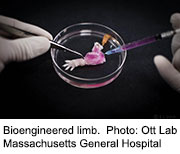


 [F1]Potential side effects

 [F2]chemical compound possessing both hydrophilic (water-loving, polar) and lipophilic (fat-loving) properties.


TOP B

********************************************************************************************************************************************

TOP C

Home


Script of the Show June  27 2015

3D Printed Cell Technique Allows Material To Mimic Human-Like Tissue

Biomimetic Technologies Project Will Create First Soft-Bodied Robots

World’s first artificial enzymes created using synthetic biology

Breakthrough in synthetic enzymes could lead to the manufacture of organisms

Synthetic enzymes hint at life without DNA or RNA

Scientists unveil giant leap towards synthetic life

Scientists create 'alien' life form with artificial genetic code

***********************************************************************************************************************************************

3D Printed Cell Technique Allows Material To Mimic Human-Like Tissue

A team of chemists has found a way to print 3D material that resembles living tissue.-The scientists developed a 3D printer that squeezes out tiny water droplets into a drop of oil, creating miniature spheres with a lipid bilayer that mimics the membranes of living cells. They deposited these droplets in layers to create a material that can bend like muscle or communicate like neurons, as reported today (April 4) in the journal Science.--"We've made a new kind of material. Not just a new material, but really a whole class of materials," said study co-author Gabriel Villar, a chemist at the University of Oxford, in England. In principle, these materials could be used to mimic any kind of living tissue[F1] , Villar told LiveScience.--The material consists of tens of thousands of water droplets separated by a thin membrane called a lipid bilayer, the same membrane that encapsulates living cells and keeps everything from spilling out. These bilayers consist of an oily, water-resistant layer sandwiched between two water-friendly layers.--Previous experiments have assembled droplets into lipid bilayers, but the droplets were either made by hand, or were made using techniques that couldn't control the movement of individual droplets.-Villar and colleagues have developed a 3D printer that could produce these droplets automatically. The printer uses a very thin glass nozzle to create droplets that are about 50 microns across (roughly half the width of a human hair). The printer squeezed the droplets into an oily solution that spontaneously coated each droplet in a lipid bilayer.--Next, the team showed how the printed material could be used to perform some of the functions of living tissue. For example, they printed a network of droplets that could transmit signals like tissues in the nervous system (albeit on a much simpler level). Neurons conduct electrical pulses along cell membranes by letting charged particles called ions pass through them. To mimic this, the researchers added a protein made by bacteria to some of the droplets, which cuts holes in lipid bilayers akin to a "cookie cutter," Villar said. An electrical current applied to the material passed through these holes much like a nerve impulse.--But the scientists didn't stop there. They used their printer to make self-folding structures, like flattened flower whose petals folded up. [See Video of Self-folding Droplets in Action]--The folding was a completely passive process that relied on the movement of water between droplets. The researchers added salt to some of the lipid-enclosed droplets, which created a salt gradient with nearby droplets. Water naturally moved from the droplets with no salt to those with salt, causing the salty droplets to swell. As a result, the flat petals curled up to form a hollow ball. The material was lifting itself against gravity, much like muscle tissue.--Ultimately, these 3D-printed materials could be used to deliver medicine or replace damaged tissue itself, the researchers said.

"This is an exciting new development in 3D printing using synthetic mimics of cells," said biomedical engineer Will Shu of Heriot-Watt University in the U.K., who was not involved in the study. "It is not hard to imagine this approach could be integrated for living organisms and the potential applications would be enormous."

****************************************************************************

Biomimetic Technologies Project Will Create First Soft-Bodied Robots

January 25th, 2007 in Technology / Engineering

A group of researchers at Tufts University has launched a multidisciplinary initiative focused on the science and engineering of a new class of robots that are completely soft-bodied. These devices will make possible advances in such far flung arenas as medicine and space exploration. Barry Trimmer, professor of biology, and David Kaplan, professor of biomedical engineering, are co-directors of the Biomimetic Technologies for Soft-bodied Robots project, which represents a consortium of seven Tufts faculty members from five departments in the School of Engineering and the School of Arts and Sciences. The project has just been awarded a grant of $730,000 from the W.M. Keck Foundation.--According to Kaplan, the project will bring together biology, bioengineering and micro/nano fabrication. “Our overall goal is to develop systems and devices--soft-bodied robots--based on biological materials and on the adaptive mechanisms found in living cells, tissues and whole organisms,” he explains. These devices, he notes, will have direct applications in robotics, such as manufacturing, emergency search and retrieval, and repair and maintenance of equipment in space; in medical diagnosis and treatment, including endoscopy, remote surgery, and prostheses design; and in novel electronics such as soft circuits and power supplies.--“A major characteristic that distinguishes man-made structures from biological ones is the preponderance of stiff materials,” explains Trimmer. “In contrast, living systems may contain stiff materials such as bone and cuticle but their fundamental building blocks are soft and elastic. This distinction between biological and man-made objects is so pervasive that our evaluation of artificial or living structures is often made on the basis of the materials alone. Many machines incorporate flexible materials at their joints and can be tremendously fast, strong and powerful, but there is no current technology that can match the performance of an animal moving through natural terrain.”

First “Molecules to Robots” Effort

The Tufts team represents the first major effort to design a truly soft-bodied locomoting robot with the workspace capabilities similar to those of a living animal. While other groups around the world are applying biomimetic approaches to engineering design, most focus on narrow areas within this field. --"This represents a wonderfully rich and novel collaboration that takes a comprehensive 'molecules to robots' approach to the use of soft materials," notes Linda M. Abriola, dean of the Tufts School of Engineering.

Work will focus on four primary areas: Control systems for soft-bodied robots, biomimetic and bionic materials, robot design and construction, and development and application of research-based platform technologies. [F2] 

Caterpillars and Silkworms

The Keck grant will provide the team with specialized equipment for use with soft materials and biomechanics experiments, according to Trimmer, whose work with caterpillars provides insights on how to build the world’s first soft-bodied robot. Trimmer, a neurobiologist, has been studying the nervous system and biology since 1990 through grants from the National Institutes of Health and the National Science Foundation. His goal has been to better understand how the creatures can control their fluid movements using a simple brain and how they can move so flexibly without any joints. He hopes to adapt his caterpillar research to this new project using the expertise of Tufts engineers.

Kaplan, whose laboratory focuses on biopolymer engineering, has already uncovered the secret of how spiders and silkworms are able to spin webs and cocoons made of incredibly strong yet flexible fibers. More recently, his team applied genetic engineering and nanotechnology to create a “fusion protein” that for the first time combined the toughness of spider silk with the intricate structure of silica. Kaplan notes that there has been tremendous progress in the development and use of soft materials in devices ranging from keyboards to toys. “However, it is very hard to make soft devices that move around and can be precisely controlled,” he says. “This is the fundamental reason why robots currently move like robots instead of lifelike animals.” --The new robots developed at Tufts will be continuously deformable and capable of collapsing and crumpling into small volumes. They will have capabilities that are not currently available in single machines including climbing textured surfaces and irregular objects, crawling along ropes and wires, or burrowing into complex confined spaces[F3] . “Soft-bodied robots could make many dangerous surgeries much safer and less painful,” Trimmer adds. “They could also be used by NASA to repair space stations by reaching places that astronauts can’t, perform more complicated tasks in industry that require flexibility of movement, help in hazardous environments like nuclear reactors and landmine detection, and squeeze more efficiently into tight spaces.”--In addition to Trimmer and Kaplan, Assistant Professors Robert White, mechanical engineering, and Sameer Sonkusale, electrical and computer engineering, will supervise projects in the Tufts Microfabrication Laboratory. Associate Professor Luis Dorfmann, civil and environmental engineering, and Visiting Assistant Professor Gary Leisk, mechanical engineering, will supervise the material testing and modeling parts of the project, and Assistant Professor Valencia Joyner, electrical and computer engineering and Sonkusale will direct the design and production of sensors and soft material integrated circuits.

Source: Tufts University

 *******************************************************************************************************************************************


World’s first artificial enzymes created using synthetic biology -

Xeno nucleic acid (XNA) is a synthetic alternative to the natural nucleic acids DNA and RNA as information-storing biopolymers that differs in the sugar backbone.[1] As of 2011, at least six types of synthetic sugars have been shown to form nucleic acid backbones that can store and retrieve genetic information. Research is now being done to create synthetic polymerases to transform XNA. The study of its production and application has created a field known as xenobiology.--Although the genetic information is still stored in the four canonical base pairs (unlike other nucleic acid analogues), natural DNA polymerases cannot read and duplicate this information. Thus the genetic information stored in XNA is “invisible” and therefore useless to natural DNA-based organisms.[2]

 http://www.cam.ac.uk/research/news/worlds-first-artificial-enzymes-created-using-synthetic-biology#sthash.Hi3SOWu0.dpuf

 Enzymes made from artificial molecules which do not occur anywhere in nature have been shown to trigger chemical reactions in the lab, challenging existing views about the conditions that are needed to enable life to happen.--Our assumptions about what is required for biological processes – the ‘secret of life’ – may need some further revision

Alex Taylor --A team of researchers have created the world’s first enzymes made from artificial genetic material.-The synthetic enzymes, which are made from molecules that do not occur anywhere in nature, are capable of triggering chemical reactions in the lab.--The research is published in the journal Nature and promises to offer new insights into the origins of life, as well as providing a potential starting point for an entirely new generation of drugs and diagnostics.[F1]  In addition, the authors speculate that the study increases the range of planets that could potentially host life.--All life on Earth depends on the chemical transformations that enable cellular function and the performance of basic tasks, from digesting food to making DNA. These are powered by naturally-occurring enzymes which operate as catalysts, kick-starting the process and enabling such reactions to happen at the necessary rate.--For the first time, however, the research shows that these natural biomolecules may not be the only option, and that artificial enzymes could also be used to power the reactions that enable life to occur.--The findings build on previous work in which the scientists, from the MRC Laboratory of Molecular Biology in Cambridge and the University of Cambridge, created synthetic molecules called “XNAs”. These are entirely artificial genetic systems that can store and pass on genetic information in a manner similar to DNA.

Using these XNAs as building blocks, the new research involved the creation of so-called “XNAzymes”. Like naturally occurring enzymes, these are capable of powering simple biochemical reactions.--Dr Alex Taylor, a Post-doctoral Researcher at St John’s College, University of Cambridge, who is based at the MRC Laboratory and was the study’s lead author, said: “The chemical building blocks that we used in this study are not naturally-occurring on Earth[F2] , and must be synthesised in the lab. This research shows us that our assumptions about what is required for biological processes – the ‘secret of life’ – may need some further revision. The results imply that our chemistry, of DNA, RNA and proteins, may not be special and that there may be a vast range of alternative chemistries that could make life possible.--Every one of our cells contains thousands of different enzymes, many of which are proteins. In addition, however, nucleic acids – DNA and its close chemical cousin, RNA – can also form enzymes. The ribosome, the molecular machine which manufactures proteins within all cells, is an RNA enzyme. Life itself is widely thought to have begun with the emergence of a self-copying RNA enzyme.--Dr Philipp Holliger, from the MRC Laboratory of Molecular Biology, said: “Until recently it was thought that DNA and RNA were the only molecules that could store genetic information and, together with proteins, the only biomolecules able to form enzymes.”“Our work suggests that, in principle, there are a number of possible alternatives to nature’s molecules that will support the catalytic processes required for life. Life’s ‘choice’ of RNA and DNA may just be an accident of prehistoric chemistry.”“The creation of synthetic DNA, and now enzymes, from building blocks that don’t exist in nature also raises the possibility that if there is life on other planets it may have sprung up from an entirely different set of molecules, and widens the possible number of planets that might be able to host life.”--The group’s previous study, carried out in 2012, showed that six alternative molecules, called XNAs, could store genetic information and evolve through natural selection. Expanding on that principle, the new research identified, for the first time, four different types of synthetic catalyst formed from these entirely unnatural building blocks.-These XNAzymes are capable of catalysing simple reactions, like cutting and joining strands of RNA in a test tube. One of the XNAzymes can even join strands together, which represents one of the first steps towards creating a living system.[F3]  Because their XNAzymes are much more stable than naturally occurring enzymes, the scientists believe that they could be particularly useful in developing new therapies for a range of diseases, including cancers and viral infections, which exploit the body’s natural processes.---Dr Holliger added: “Our XNAs are chemically extremely robust and, because they do not occur in nature, they are not recognised by the body’s natural degrading enzymes[F4] . This might make them an attractive candidate for long-lasting treatments that can disrupt disease-related RNAs.”---Professor Patrick Maxwell, Chair of the MRC’s Molecular and Cellular Medicine Board and Regius Professor of Physic at the University of Cambridge, said: “Synthetic biology is delivering some truly amazing advances that promise to change the way we understand and treat disease. The UK excels in this field, and this latest advance offers the tantalising prospect of using designer biological parts as a starting point for an entirely new class of therapies and diagnostic tools that are more effective and have a longer shelf-life.”-Funders of the research included the MRC, European Science Foundation and the Biotechnology and Biological Sciences Research Council.The text in this work is licensed under a Creative Commons Licence. If you use this content on your site please link back to this page. For image rights, please see the credits associated with each individual image.- See more at: http://www.cam.ac.uk/research/news/worlds-first-artificial-enzymes-created-using-synthetic-biology#sthash.Hi3SOWu0.dpuf

 ******************************************************************

 Breakthrough in synthetic enzymes could lead to the manufacture of organisms

  • Synthetic form of DNA, called XNA, is capable of editing genetic material
  • XNA triggered reactions thought to be crucial for life first starting on Earth
  • Scientists suggest alien life could have evolved using XNA instead of DNA
  • Artificial molecules could also be used to create synthetic life in the lab
  • Researchers believe XNA may lead to a new ways of treating cancers


The world’s first enzymes made from artificial genetic material have been created by scientists in what could be a major step towards generating synthetic life.-The enzymes, which do not occur naturally, were created using a synthetic form of DNA called XNA and were capable of triggering chemical reactions in the lab.-The findings build on previous work that showed six types of XNA molecules were capable of storing and transmitting genetic information in the same way as DNA and RNA.-It had been previously thought that DNA and RNA, which form the basis for all life on Earth, were the only way of storing genetic material.-But now the latest research by synthetic biologists in Cambridge shows that this synthetic genetic material is also capable of performing another crucial biological role - catalysing biochemical reactions that are essential for life.-Using their lab-made XNAs as building blocks, the team were able to create synthetic enzymes, which they have named ‘XNAzymes’, that could cut up and stitch together small chunks of genetic material, just like naturally occurring enzymes.-The suggests that such molecules could be used to replicate some of the earliest steps needed to produce life itself and may even provide clues about what life on other planets may be like.-It is thought that life first began with the evolution of a segment of RNA that was able to copy itself and catalyse reactions. If XNA is also capable of this, then it could also have led to different forms of life on other planets or could be used to create new synthetic forms of life. However, Dr Philipp Holliger, who led the research at the MRC Laboratory of Molecular Biology in Cambridge, said: ‘Our work suggests that, in principle, there are a number of possible alternatives to nature's molecules that will support the catalytic processes required for life.-‘Until recently, it was thought that DNA and RNA were the only molecules that could store genetic information and, together with proteins, the only biomolecules able to form enzymes.-‘Life's 'choice' of RNA and DNA may just be an accident of prehistoric chemistry.--'The creation of synthetic DNA, and now enzymes, from building blocks that don't exist in nature also raises the possibility that if there is life on other planets it may have sprung up from an entirely different set of molecules, and widens the possible number of planets that might be able to host life.’-In 2012 Dr Holliger’s group showed that there were six alternative molecules to the oligonucleotides that form RNA and DNA, which they called XNAs.--They demonstrated that these could store information and could even evolve through natural[F5]  selection.-In their latest research, which is published in the journal Nature, the team created four different types of synthetic enzyme from strands of XNA.--These XNAzymes were able to perform the role of a polymerase - an enzyme that cuts and joins RNA strands together - in a test tube. One of the XNAzymes they created was also able to join XNA strands together to form longer molecules - a key step towards creating a living system that can replicate itself.--[F6] Although it will still be some time before these can be used to create living synthetic organisms, Dr Holliger believes that XNAzymes could also be useful for developing new therapies for range of diseases including cancers and some viral infections.[F7] -Dr Holliger added: ‘Our XNAs are chemically extremely robust and, because they do not occur in nature, they are not recognised by the body's natural degrading enzymes.-[F8] ‘This might make them an attractive candidate for long-lasting treatments that can disrupt disease-related RNAs.’Professor Patrick Maxwell, chair of the MRC's Molecular and Cellular Medicine Board, said the work could kick start an entirely new branch of medicine.One of the XNA molecules created at the Laboratory of Molecular Biology in Cambridge which was capable of joining two strands of XNA together

The XNAzymes created by the scientists were also able to cut up strands of RNA, another crucial biochemical reaction that is thought to have been fundamental in kick starting life on Earth

Two of the XNA enzymes created by the scientists at the Laboratory of Molecular Biology in Cambridge, which were able to join two strands of XNA together (left) and cut up strands of RNA (right)-Life on other planets outside our own solar system, like this exoplanet, could have evolved from XNA rather than RNA, which could have led to very different forms of life to those we are familiar with here on Earth--He said: ‘Synthetic biology is delivering some truly amazing advances that promise to change the way we understand and treat disease.[F9]  ‘This latest advance offers the tantalising prospect of using designer biological parts as a starting point for an entirely new class of therapies and diagnostic tools that are more effective and have a longer shelf-life.’-Professor Jack Szostak, a Nobel prize winner at Harvard University who studies the origins of life, added that the research raises some fundamental questions about what life on other planets may be like.He told New Scientist: ‘The possibility that life elsewhere, on exoplanets, could have started with something other than RNA or DNA is quite interesting. ‘But the primordial biopolymer for any form of life must satisfy other constraints as well, such as being something that can be generated by prebiotic chemistry and replicated efficiently.-‘Whether XNA can satisfy these constraints, as well as providing useful functions, remains an open question.’ 

**********************************************************************
 
Synthetic enzymes hint at life without DNA or RNA

Life might not have to be based on DNA or RNA

Enzymes that don't exist in nature have been made from genetic material that doesn't exist in nature either, called XNA, or xeno nucleic acid.--It's the first time this has been done and the results reinforce the possibility that life could evolve without DNA or RNA, the two self-replicating molecules considered indispensible for life on Earth.--"Our work with XNA shows that there's no fundamental imperative for RNA and DNA to be prerequisites for life," says Philipp Holliger of the Laboratory of Molecular Biology in Cambridge, UK, the same laboratory where the structure of DNA was discovered in 1953 by Francis Crick and James Watson.

It's not all about the base--

Holliger's team has made XNAs before. Their unnatural XNA contains the same bases – adenine, thymine, guanine, cytosine and uracil – on which DNA and RNA rely for coding hereditary information. What's different is the sugar to which each base is attached.--In DNA and RNA, the sugars are deoxyribose and ribose, respectively. Holliger made new types of genetic material by replacing these with different sugars or other molecules.--Now, they have taken a step closer to mimicking early life on the planet by showing that XNAs can also serve as enzymes – indispensible catalysts for speeding up chemical reactions vital for life.--One of the first steps towards life on Earth is thought to be the evolution of RNA into self-copying enzymes.

Big steps

So by showing that XNAs can act as enzymes, on top of being able to store hereditary information, Holliger has recreated a second major step towards life.--The XNA enzymes can't yet copy themselves but they can cut and paste RNA, just like natural enzymes do, and even paste together fragments of XNA.--It's the first demonstration that, like prehistoric RNA, XNA can catalyse reactions on itself, even if it can't yet copy itself as RNA can.--Holliger argues that RNA and DNA may have come to dominate Earth by chance, simply because they were the best evolutionary materials to hand. "You could speculate that on other planets, XNAs would dominate instead," he says.

Primal molecules

"This work is another nice step towards demonstrating the functional capabilities of XNAs," says Nobel prizewinner Jack Szostak of Harvard University, who studies the origins of life on Earth .--"The possibility that life elsewhere, on exoplanets, could have started with something other than RNA or DNA is quite interesting, but the primordial biopolymer for any form of life must satisfy other constraints as well, such as being something that can be generated by prebiotic chemistry and replicated efficiently," Szostak says. "Whether XNA can satisfy these constraints, as well as providing useful functions, remains an open question."

Holliger says that XNAs may also have roles to play in medicine. Because they do not occur naturally, they can't be broken down in the human body. And since they can be designed to break and destroy RNA, they could work as drugs for treating RNA viruses or disabling RNA messages that trigger cancers.-"We've made XNA enzymes that cut RNA at specific sites, so you could make therapies for cleaving viral or oncogenic messenger RNA[F10] ," says Holliger. "And because they can't be degraded, they could give long-lasting protection."

Journal reference: Nature, DOI: 10.1038/nature13982

*******************************************************************

Scientists unveil giant leap towards synthetic life

Achievement akin to ‘climbing Mount Everest’ in its complexity

Friday, 28 March 2014

Scientists have made the first artificial chromosome which is both complete and functional in a milestone development in synthetic biology, which promises to revolutionise medical and industrial biotechnology in the coming century.--The researchers built the artificial chromosome[F11]  from scratch by stitching synthetic strands of DNA together in a sequence based on the known genome of brewer’s yeast. They predict that a completely synthetic yeast genome comprised of its entire complement of 16 chromosomes could be made within four years.-“Our research moves the needle in synthetic biology from theory to reality. This work represents the biggest step yet in an international effort to construct the full genome of synthetic yeast,” said Jef Boeke of the New York University School of Medicine, a lead author of the study published in the journal Science.-“It is the most extensively altered chromosome ever built. But the milestone that really counts is integrating it into a living yeast cell. We have shown that yeast cells carrying this synthetic chromosome are remarkably normal,” [F12] Dr Boeke said.--“They behave almost identically to wild yeast cells, only they now possess new capabilities and can do things that wild yeast cannot [do],” he said.--“Not only can we make designer changes on a computer, but we can make hundreds of changes through a chromosome and we can put that chromosome into yeast and have a yeast that looks, smells and behaves like a regular yeast, but this yeast is endowed with special properties that normal yeasts don’t have,[F13] ” he explained.--The synthetic yeast chromosome was based on chromosome number 3, but scientists deleted large parts of it that were considered redundant and introduced further subtle changes to its sequence – yet the chromosome still functioned normally and replicated itself in living yeast cells, they said.--“We took tiny snippets of synthetic DNA and fused them together in a complex series of steps to build an essentially computer-designed chromosome 3, one of the 16 chromosomes of yeast. We call it ‘synIII’ because it’s a completely synthetic derivative that has been engineered in a variety of interesting ways to make it different from the normal chromosome,” Dr Boeke said.--The achievement was compared to climbing Mount Everest in its labour-intensive complexity, as it involved stitching together 273,871 individual building blocks of DNA – the nucleotide bases of the yeast’s genes – in the right order, and removing about 50,000 repeating sequences of the chromosome that were considered redundant.--“When you change the genome you’re gambling. One wrong change can kill the cell. We have made over 50,000 changes to the DNA code in the chromosome and our yeast still lived. That is remarkable, it shows that our synthetic chromosome is hardy, and it endows the yeast with new properties,” Dr Boeke said.--Britain is one of several countries involved in the international effort to synthesise all 16 yeast chromosomes. Last year, the Government announced that it will spend £1 million on the yeast project out of a total budget of £60 million it has dedicated to synthetic biology.--Paul Freemont of Imperial College London said that the first complete and functional synthetic yeast chromosome is “a big deal” and significant step forward from the work by DNA scientist Craig Venter, who synthesised the much simpler genome of a bacterium in 2010.--“It opens up a whole new way of thinking about chromosome and genome engineering as it provides a proof of concept that complicated chromosomes can be redesigned, synthesised and made to work in a living cell,” Dr Freemont said.--Artificial chromosomes designed by computer will be vital for the synthetic life-forms that scientists hope to design for a range of applications, such as the breakdown of persistent pollutants in the environment or the industrial manufacture of new kinds of drugs and vaccines for human and animal medicine.--“It could have a lot of practical applications because yeast is used in the biotechnology industry to produce everything from alcohol, which has been produced for centuries, to biofuels and speciality chemicals to nutrients,” Dr Boeke said.--“Yeast is a really interesting microorganism to work on because it has an ancient industrial relationship with man. We’ve domesticated it since the days of the Fertile Crescent and we’ve had this fantastic collaboration to make wine, break and beer,” he said.--“That relationship persists today in a wide range of products that are made with yeast such as vaccines, fuels and specialty chemicals and it’s only going to be growing. Yeast is one of the few microbes that packages its genetic material in a nucleus just like human cells. So it serves as a better model for how human cells work in health and disease,” Dr Boeke added.


.

**********************************************************************

Scientists create 'alien' life form with artificial genetic code


From left to right, the structures of A-, B- and Z-DNA. Zephyris

Scientists made a substantial breakthrough in understanding how to alter the fundamental nature of life, and they did so by creating for the first time a partially artificial life form that passes along lab-engineered DNA. --The work, published online in the journal Nature on Wednesday, came from the Scripps Research Institute in La Jolla, Calif., and centered around a modified strain of E. coli bacterium that was fused with chemically synthesized nucleotides and was able to replicate its natural and synthetic components during reproduction[F14] . -Throughout the entire history of life on Earth, the genetic code of all organisms has been uniform, from the simplest of bacteria all the way up to human beings, meaning our genetic code is composed of the same four nucleotides labeled A, C, T, and G. Those nucleotides join to form base pairs, which are used in the creation of genes that cells use to produce proteins. --Researchers at Scripps created two new nucleotides, X and Y, and fused them into the E. coli bacterium. The organism was able to reproduce normally with six -- instead of the standard four -- nucleotides, meaning it genetically passed along the first combination of manmade and natural DNA. -"This has very important implications for our understanding of life," Floyd Romesberg, who headed the Scripps researcher team, told The New York Times. "For so long people have thought that DNA was the way it was because it had to be, that it was somehow the perfect molecule." --Because this breakthrough could impact more than just biological research, the field -- called synthetic biology -- is likely to be met with harsh criticism from those who fear that tampering with the building blocks of existence could be a step too far for science. The subset of synthetic biology focusing on creations unfamiliar to nature with expanded genetic alphabets is sometimes referred to as xenobiology. --"The arrival of this unprecedented 'alien' life form [F15] could in time have far-reaching ethical, legal and regulatory implications," Jim Thomas of the ETC Group, a Canadian advocacy organization, told The New York Times. "While synthetic biologists invent new ways to monkey with the fundamentals of life, governments haven't even been able to cobble together the basics of oversight, assessment or regulation for this surging field."[F16] -To create a modified organism that would reproduce, Romesberg's team had to first create stable enough artificial nucleotides. The creation of X and Y variants came only after 300 types were tried. The X nucleotide pairs with the Y, just as A does with T and C with G in natural DNA. It's unclear whether a semi-artificial organism could sustain a far more expansive genetic code, meaning many more synethic pairs, and if there is any time-based restraint involved.-As far as worrying about never-before-seen strains of bacteria escaping into the wild, Romesberg stressed that this newly created organism could never infect anything[F17] . To continue reproducing the synthetic nucleotides, the researchers had to feed the necessary chemicals to the bacterium or else it would stop producing the X and Y pair. P--Romesberg and his colleagues' findings follow decades of work in synthetic biology, and the results have long since left the confines of academic research. Romesberg's company, Synthorx, is trying to design an administering technique for viruses that would rely on the artificial life forms' inability to reproduce the synthetic nucleotides without the proper chemicals, meaning they could be used to create an immune system response while be inhibited from spreading.

Beyond those immediate applications, the next steps are figuring out if the synthesized nucleotides can be fused into the RNA of living organisms and used to produce new proteins, as well as discovering whether or not genetically engineered cells could be used to help organisms reproduce those synthetic nucleotides on their own.


 [F1]The paradym has changed even in the health in alternatives this will change everything---this DNA or rather XNA is environmental will alter the natural things and alter them in ways  we cannot conceive-which will also alter us and furthering some genetic change or alterations and possibly mutations in the physiology that can produce unknown anomalies or conditions or even furthering the life span to  either become less or more or potentially disrupt the immune system to make it easier to afflict or potentially augment the immune system—at this point no clarity but to design a drug with an unknown DNA –could  be irreversible

 [F2]So are they from the space station up in orbit—are they from some parallel-or are they even alien? It is a thought provoker~where ?

 [F3]How morgellons or nanopoisoningwould be made possible

 [F4]Simplest layman’s terms no defence against this—with out enzymes to break them down and since they are a synthetic DNA they could very easily over write the normal DNA to cause mutations to occur

 [F5]Imagine that New XNA that could evolve through Natural Selection---what happens to DNA??

 [F6]Excite me Already---creating life with a XNA no one knows really anything about---and does XNA produce a X-MAN or X-WOMAN!!!

 [F7]I love this`` another justification on this is going to be Good for mankind when all they have said leads one to think the opposite a new  means to inflict-afflict or terminate

 [F8]And this is comforting How? The y do not degrade in the system because they are not recognized –so that means sine the system is not aware of this XNA it will go without any check or balance to reduce this when it is finished –when it is done it’s job it may then stick around and adapt to or attach itself to DNA orrrr possibly replicate itself in a host and replace the DNA eventually---this is called mutation

 [F9]A more effective way for war---the best way to treat and define diseases is to eliminate the weak or afflicted---case resolved XNA

 [F10]Doesn’t this make one wonder why there has not been a cure when you can add XNA enzyme to converge with DNA and perpuate a condition indefinitely since the DNA has no enzyme to break it down---perfect pathogen to barely keep one aive and support the perpetual entanglement of a pharma stronghold

 [F11]Chromosomes are thread-like structures located inside the nucleus of animal and plant cells. Each chromosome is made of protein and a single molecule of deoxyribonucleic acid (DNA). Passed from parents to offspring, DNA contains the specific instructions that make each type of living creature unique

 [F12]And so many people are filled with yeast—and can’t seem to get rid of it—maybe there us a connection

 [F13]Would we call this Genetic Engineering—Artificial Life-or a BioNano tech—Or a weapon

 [F14]Would call this Gentically modified Bioweapons

 [F15]They are slyly telling us what it really is in this statement—truth right in the open

 [F16]This is such BS—Gov’t have never came up with anyway to regulate anything in Biotech---that is alarmingly ridiculous---the ones who dictate the legislation has always been the oes who supported the funds for these projects such as Monsanto-symbus-bayer-dupont-basf Pfizer..etc—these are the ones whowill dictate policy and the gov’t will obey not the other way around

 [F17]This is another one of those mystical magical statement of how inert it is but with all the interaction they are saying the YNA can do it would be a catastrophe if it got out


 [F1]Would appear to be a construct of  AL since these would compliment a XNA—living tissue does not refer to human living or Animal

 [F2]Synthetic Life or Artificial Life

 [F3]Insect like qualities


TOP C